Fig. 1
- ID
- ZDB-FIG-080424-43
- Publication
- Novince et al., 2003 - Cadherin expression in the inner ear of developing zebrafish
- Other Figures
- All Figure Page
- Back to All Figure Page
|
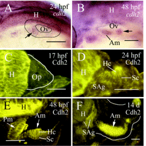
Expression of Cdh2 in the inner ear of developing zebrafish. Panels (A) and (B) are lateral views of the otic vesicle (Ov) of 24 hpf and 48 hpf whole-mount embryos, respectively, labeled by in situ hybridization with cadherin-2 cRNA (cdh2). Dorsal is up and anterior is to the left. Panels (C–F) are cross sections of the inner ear stained with Cadherin-2 antibody (Cdh2). The arrow in panel (A) points to a sensory patch in the anterioventral portion of the otic vesicle. The arrow in panel (B) indicates a cdh2-positive neuromast in the epidermis above the inner ear. Panel (F) is a surface view of the anterior macula (Am). Other abbreviations: H, hindbrain; Hc, hair cells; Pm, posteriomedial macula; Op, otic placode; SAg, statoacoustic ganglion; Sc, supporting cells. Scale bar: 50 μm. |
Reprinted from Gene expression patterns : GEP, 3(3), Novince, Z.M., Azodi, E., Marrs, J.A., Raymond, P.A., and Liu, Q., Cadherin expression in the inner ear of developing zebrafish, 337-339, Copyright (2003) with permission from Elsevier. Full text @ Gene Expr. Patterns

