
Fig. 3
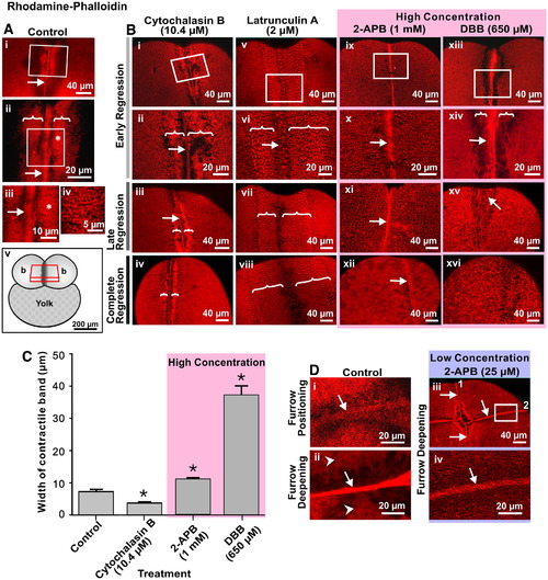
Intracellular Ca2+ is required for actin remodeling during cytokinesis. Representative top views of fixed embryos labeled with rhodamine–phalloidin that were (A) untreated (control) or (B) treated with 10.4 μM cytochalasin B; 2 μM Latrunculin A; 1 mM 2-APB or 650 μM DBB. In the case of the control (A), images are shown in the region of the first cleavage furrow during late deepening. Panels Aii and Aiii are magnified views of the white boxes marked on panels Ai and Aii, respectively. Panel iv is a magnified view of the region shown with an asterisk in panel Aiii. White arrows and brackets indicate the contractile band and the PAEs, respectively. The schematic (panel Av) indicates the location within the embryo of the stack of 2-D image sections used to reconstruct the images shown in A and B. (B) In the case of the various drug treatments, images were acquired during early regression (panels i, ii, v, vi, ix, x, xiii and xiv), late regression (panels iii, vii, xi and xv) and on complete regression (panels iv, viii, xii and xiv). Panels ii, vi, x and xiv are magnified views of the white boxes marked on panels i, v, ix and xiii, respectively. Again, the white arrows and brackets indicate the contractile bands and the PAEs, respectively. (C) Measurement of the contractile band width in cytochalasin B, 2-APB and DBB-treated embryos during early furrow regression. Treatment with cytochalasin B had a significant effect on reducing the width of the contractile band compared to the control, whereas 2-APB and to a much greater degree, DBB, had the opposite effect and significantly increased the width of the band. Data presented are averaged values ± S.E.M. with n = 3 to 5. * indicates values of contractile band width that are significantly different from the control (at p < 0.05 for the CB- and 2-APB-treated embryos, and p < 0.001 for the DBB-treated embryos). (D) Actin remodeling defects in embryos treated with a low concentration of 2-APB. Representative top views of fixed embryos labeled with rhodamine–phalloidin during: furrow positioning (panel i) and furrow deepening (panel ii) of the second cleavage in untreated (control) embryos; and at the end of the second cleavage (panels iii and iv) following treatment with 25 μM 2-APB shortly after the appearance of the first cleavage furrow. Panel iv is a magnified view of the white box in panel iii. In panels i and ii, the arrows indicate the actin rafts and compact contractile band, respectively, which occur during normal cleavage, while the arrowheads in panel ii indicate the PAEs on either side of the contractile band. In panels iii and iv, the numbers (1 and 2) indicate the first and second cleavage furrows, while the arrows indicate the abnormal contractile bands that are visible in the blastoderm cortex following low-concentration 2-APB treatment. |
Reprinted from Developmental Biology, 316(2), Li, W.M., Webb, S.E., Chan, C.M., and Miller, A.L., Multiple roles of the furrow deepening Ca2+ transient during cytokinesis in zebrafish embryos, 228-248, Copyright (2008) with permission from Elsevier. Full text @ Dev. Biol.