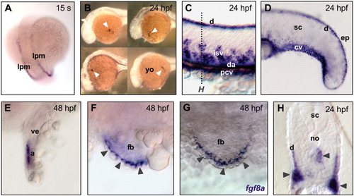
Whole-mount in situ hybridization for emilin2a in zebrafish embryos at different developmental stages. A: Expression in cells of the lateral mesoderm at 15 somites (s). B: Expression by cells in the yolk, corresponding to macrophage clusters (arrowheads), at 24 hours postfertilization (hpf). C,D: Expression in the circulatory system and in the dermis of trunk (C) and tail (D) at 24 hpf. E: Expression in the atrium of the developing heart at 24 hpf. F: Expression in the apical ectodermal ridge (arrowheads) of fin buds at 48 hpf. G: Staining of the apical ectodermal ridge (arrowheads) of fin buds with a fgf8a probe at 48 hpf, showing the same pattern of emilin2a. H: Cross-section of a 24 hpf embryo (level of section is marked by a dotted line in C), showing expression in the vasculature (arrowheads) and in the dermis. With the exception of A and B, the yolk was removed and embryos were flattened. A is a dorsal view, anterior side of embryo is at the top; B-G are lateral views, anterior side is on the left; in the cross-section, dorsal side is at the top. a, atrium; cv, caudal vein; d, dermis; da, dorsal aorta; ep, epidermis; fb, fin bud; isv, intersegmental blood vessels; lpm, lateral plate mesoderm; no, notochord; pcv, posterior cardinal vein; sc, spinal cord; ve, heart ventricle; yo, yolk.
|
