
Fig. 4
- ID
- ZDB-FIG-080325-63
- Publication
- Wilkins et al., 2008 - Mtx2 directs zebrafish morphogenetic movements during epiboly by regulating microfilament formation
- Other Figures
- All Figure Page
- Back to All Figure Page
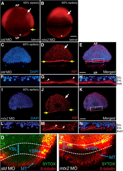
Cytoskeletal elements of the zebrafish yolk during epiboly. (A, B) Rhodamine–phalloidin (RP) stained embryos at 60% epiboly, pre-injected with std MO (A) or mtx2 MO (B). RP stains the (vegetal) actin mat (asterisks), the F-actin ring of the E-YSL (yellow arrow in panel A only) and peripheral F-actin within EVL cells (white arrows). (C–N) Confocal analysis of RP (D, G, J, M) and DAPI (C, F, I, L) stained 60% epiboly stage embryos. (G–L) std MO embryos have a punctate F-actin ring, vegetal to the blastoderm margin (yellow arrows in panel D). Detail of the dotted box in panel E is shown in panels F–H. (I–N) mtx2 MO injected embryos also stain the periphery of EVL cells (white arrows in panels D, J), the leading edge of the EVL (white arrowheads in panels G, M), but not the punctate F-actin ring (yellow arrows in panel J). In std MO controls the larger DAPI stained nuclei (in panels F, H) are EVL nuclei that have migrated past the, smaller, deep layer nuclei. These nuclei are less obvious in mtx2 MO embryos (L, N). nel J). In std MO controls the larger DAPI stained nuclei (in panels F, H) are EVL nuclei that have migrated past the, sma(O, P) Confocal image projection of microtubule organization at 70% epiboly (β-tubulin immunofluorescence). A microtubule network is present in both std and mtx2 MO injected embryos (arrow in panels O and P). All nuclei (including YSN) were counterstained with SYTOX Green (arrowhead in panels O and P). Overlapping tubulin/SYTOX staining patterns result in ″yellow″ fluorescence. |
| Fish: | |
|---|---|
| Knockdown Reagent: | |
| Observed In: | |
| Stage: | Shield |
Reprinted from Developmental Biology, 314(1), Wilkins, S.J., Yoong, S., Verkade, H., Mizoguchi, T., Plowman, S.J., Hancock, J.F., Kikuchi, Y., Heath, J.K., and Perkins, A.C., Mtx2 directs zebrafish morphogenetic movements during epiboly by regulating microfilament formation, 12-22, Copyright (2008) with permission from Elsevier. Full text @ Dev. Biol.