
FIGURE
Fig. 4
- ID
- ZDB-FIG-080303-8
- Publication
- Wen et al., 2008 - Visualization of monoaminergic neurons and neurotoxicity of MPTP in live transgenic zebrafish
- Other Figures
- All Figure Page
- Back to All Figure Page
Fig. 4
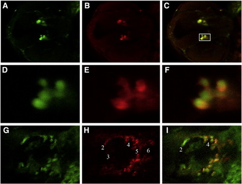
Double immunofluorescence staining for detection of GFP and endogenous Th. Simultaneous immunofluorescence staining was generated using a polyclonal anti-GFP antibody (A, D, G) and a monoclonal anti-Th antibody (B, E, H) for 30 hpf (A–F) and 3 dpf (G–I) ETvmat2:GFP embryos. Composite photos were made from stacked confocal images, focusing on catecholaminergic neurons of the ventral diencephalon. Merged pictures of Th and GFP staining are shown in panels C and F. Higher magnification images of the posterior tubercular neurons (D–F) correspond to the framed region in panel C. |
Expression Data
| Genes: | |
|---|---|
| Fish: | |
| Anatomical Term: | |
| Stage Range: | Prim-15 to Protruding-mouth |
Expression Detail
Antibody Labeling
Phenotype Data
Phenotype Detail
Acknowledgments
This image is the copyrighted work of the attributed author or publisher, and
ZFIN has permission only to display this image to its users.
Additional permissions should be obtained from the applicable author or publisher of the image.
Reprinted from Developmental Biology, 314(1), Wen, L., Wei, W., Gu, W., Huang, P., Ren, X., Zhang, Z., Zhu, Z., Lin, S., and Zhang, B., Visualization of monoaminergic neurons and neurotoxicity of MPTP in live transgenic zebrafish, 84-92, Copyright (2008) with permission from Elsevier. Full text @ Dev. Biol.