Fig. 4
- ID
- ZDB-FIG-080225-8
- Publication
- Sittaramane et al., 2008 - Expression of unconventional myosin genes during neuronal development in zebrafish
- Other Figures
- All Figure Page
- Back to All Figure Page
|
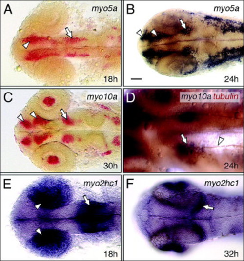
Myosin expression in the forebrain and midbrain. All panels show dorsal views of the head with anterior to the left. (A and B) At 18 hpf, myo5a is expressed in nascent neurons (arrowhead, A) in the forebrain, and in the nucMLF neurons (arrow) in the midbrain. By 24 hpf (B), forebrain expression has refined into distinguishable clusters representing the neurons of the anterior and post-optic commissures (arrowheads, B), and is maintained in the nucMLF neurons (arrow). (C) At 30 hpf, myo10a is expressed in the neurons of the nucMLF (arrow), and of the anterior and post-optic commissures (arrowheads), and in the lens (see Fig. 6). (D) Double-labeling shows that tubulin antibody-labeled axons (arrowhead) of the MLF arise from the cluster of myo10a-expressing neurons (arrow), corresponding to the nucMLF. (E and F) At 18 hpf, myo2hc1 is expressed in the medial layer of the optic cup (arrowheads), and at the midbrain–hindbrain boundary (arrow). By 32 hpf (F), expression is restricted to the retinal ganglion layer (see Fig. 6), and to the caudal edge of the developing optic tectum (arrow). Scale bar, 25 μm (D), 50 μm (A–F). |
| Genes: | |
|---|---|
| Fish: | |
| Anatomical Terms: | |
| Stage Range: | 14-19 somites to Prim-15 |
Reprinted from Gene expression patterns : GEP, 8(3), Sittaramane, V., and Chandrasekhar, A., Expression of unconventional myosin genes during neuronal development in zebrafish, 161-170, Copyright (2008) with permission from Elsevier. Full text @ Gene Expr. Patterns

