
FIGURE
Fig. 4
- ID
- ZDB-FIG-080205-5
- Publication
- Ishimaru et al., 2005 - Two families of candidate taste receptors in fishes
- Other Figures
- All Figure Page
- Back to All Figure Page
Fig. 4
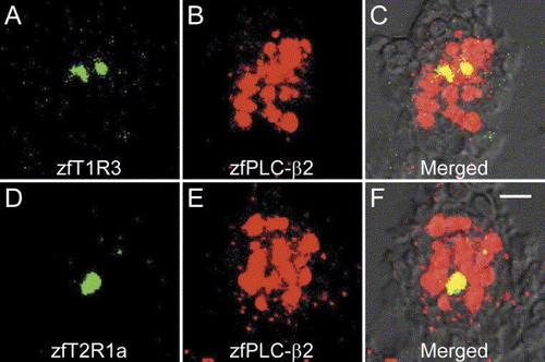
Co-expression of zfTRs with zfPLC-β2. Sections of pharyngeal region were hybridized with zfTR and zfPLC-β2 probes. The signals of probes were represented as pseudocolors (green for zfTRs and red for zfPLC-β2). The cells positive to zfT1R3 (A) or zfT2R1a (D) were included in the cells positive to zfPLC-β2 (B and E) as indicated by the yellow signals in merged images (C and F). Scale bar=10 μm. |
Expression Data
Expression Detail
Antibody Labeling
Phenotype Data
Phenotype Detail
Acknowledgments
This image is the copyrighted work of the attributed author or publisher, and
ZFIN has permission only to display this image to its users.
Additional permissions should be obtained from the applicable author or publisher of the image.
Reprinted from Mechanisms of Development, 122(12), Ishimaru, Y., Okada, S., Naito, H., Nagai, T., Yasuoka, A., Matsumoto, I., and Abe, K., Two families of candidate taste receptors in fishes, 1310-1321, Copyright (2005) with permission from Elsevier. Full text @ Mech. Dev.