
Fig. 2
- ID
- ZDB-FIG-071228-14
- Publication
- Lane et al., 2002 - Dynamic expression and regulation by Fgf8 and Pou2 of the zebrafish LIM-only gene, lmo4
- Other Figures
- All Figure Page
- Back to All Figure Page
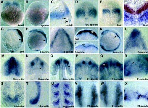
Expression of lmo4 through the 24-somite stage. Whole-mount in situ hybridization with an lmo4 probe was performed as reported (Sagerström et al., 1996). (A, B) Cleavage stages. Maternal lmo4 expression is visible at the two-cell stage (A), but is not detectable by late blastula stage (B). (C–G) Gastrula stages. (C) Lateral view of a shield stage (6 hpf) close-up showing lmo4 expression is excluded from the margin of the shield (arrow) above the forerunner cells (arrowhead). Dorsal views of (D) 75% epiboly (8 hpf), (E) bud stage (10 hpf), (F) double in situ hybridization showing lmo4 expression in blue and pax2.1 (upper stripe) and the rostral stripe (presumptive R3) of krox20 in red, in a bud stage embryo. (G) Lateral view of bud stage (10 hpf) embryos demonstrating that zygotic lmo4 is restricted to the rostralmost portion of this domain by the end of gastrulation. The arrow in (G) points to mesendodermal staining. (H, I) Three-somite stage. (H) Lateral view showing decreasing expression in the ectoderm at the three-somite stage (11 hpf), expression in the somitic mesoderm (arrowhead) and low expression in the presomitic mesoderm. (I) Animal pole view of the embryo in (H), showing expression in the anterior neural plate. (J, J2) Optical section through the rostral hindbrain area of a bud stage embryo and a six-somite stage embryo, showing expression in the neural plate (np) at bud but not six-somite stage. (K, L) Six-somite stage. (K) Neural plate expression is restricted to the anterior. Mesodermal expression persists in the somites and head mesoderm (arrow) and expression in presomitic mesoderm becomes detectable. (L) Animal pole view of embryo in (K). (M–R) Expression in the optic primordia from the ten-somite to the 21-somite stage. (M) Ten-somite stage: expression in the telencephalon (t) and much of the optic primordia. (N) Expression at the 14-somite stage is resolved to the presumptive stalk and pigmented epithelium (rpe, arrowhead). (O) Expression at the 18-somite stage in the optic stalk and rpe (arrowhead). (P) Dorsal, (Q) ventral and (R) lateral view of the optic primordia of 21-somite stage embryos. The arrowhead in (R) points to a retinal pigment cell. (S–U) Expression in somitic, presomitic and tail mesoderm. (S) Dorsal view of the tail bud presomitic mesoderm at the six-somite stage (see also (J)). (T) Lateral view of tail mesoderm at the 14-somite stage. (U) Expression in rostral somites at the 14-somite stage. (V–X) Twenty-one-somite stage. (V) Dorsal view showing expression in the otic vesicle. (W) Same embryo as in (V) in a ventral focal plane, showing expression in the pharyngeal arch mesenchyme ventral to the otic vesicle. (X) Lateral view showing expression in the anterior sensory ganglia. |
| Genes: | |
|---|---|
| Fish: | |
| Anatomical Terms: | |
| Stage Range: | 2-cell to 20-25 somites |
Reprinted from Mechanisms of Development, (Suppl.) 119, Lane, M.E., Runko, A.P., Roy, N.M., and Sagerström, C.G., Dynamic expression and regulation by Fgf8 and Pou2 of the zebrafish LIM-only gene, lmo4, S185-S189, Copyright (2002) with permission from Elsevier. Full text @ Mech. Dev.