|
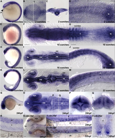
In situ hybridization using cdc25a (a-r) and cdc25d (s-v) antisense probes. Stages are indicated in the lower left of each panel. p and q are cross-sections through a 24hpf embryo at the positions labeled in l o. v is a section as indicated in t. Anatomical structures, where appropriate for orientation, are labeled. Embryo orientations are as follows: a, e, h, and k are lateral views. b, f, I, l, o, and s are dorsal views of the anterior regions of the embryo, anteriormost to the left. d, g are similarly oriented but focused on the posterior region of the embryo. j, m, n, r, t, and u are lateral views. Cross-sections c, p, q, and v are dorsal up. tb, tailbud; n, notochord; pl, polster; fb, forebrain; mb, midbrain; hb, hindbrain; ov, optic vesicle; ye, yolk extension; nt, neural tube; np, nasal placodes. Scale bar = 20 μm.
|