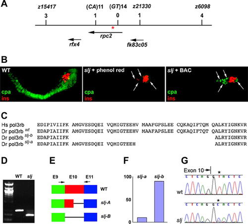
The slj Locus Encodes Zebrafish polr3b, the Gene Encoding the Second Largest Subunit of RNA Pol III (A) Schematic of the region surrounding the slj locus. Genetic markers and the number of recombinants are listed above the names of the genes and expressed sequence tags (ESTs) that map to this region of zebrafish Chromosome 18. A zero recombinant marker (GT)14, is located within the polr3b coding region. Arrows denote orientation of gene transcription. Red asterisk (*) denotes location of slj mutation. (B) High-power, lateral views of the immunostained pancreas from a 5-dpf wild-type zebrafish larva (left panel), and two slj larvae microinjected with either phenol red vehicle (middle) or the BAC spanning the slj locus (right panel). All larvae were processed for insulin (ins, red) and carboxypeptidase A (cpa, green) immunohistochemistry. A normal pattern of immunoreactive insulin is present in all larvae. BAC injection restores cpa staining and therefore partially rescues the slj exocrine (carboxypeptidase A) defect. Arrows point to cpa-positive exocrine pancreas cells in the BAC-injected larva and note their absence in the slj larva. (C) Predicted amino acid sequence encoded by the human (Hs) and zebrafish (Dr) Polr3b cDNA surrounding the region deleted by the slj mutation. Two polr3b mRNA splice variants are generated by the slj mutation: the slj-b variant lacks all 41 amino acids encoded by exon 10 (aa239–279) of the polr3b gene, whereas the slj-a variant lacks 22 exon 10 amino acids (aa258–279). (D) PCR amplification of cDNA derived from pooled 5-dpf slj larvae and homozygous wild-type (WT) siblings using primers that span polr3b exon 10, as depicted in (E). Sequencing revealed that a 278-bp band is amplified from wild-type larvae, whereas a 155-bp band corresponding to deletion of exon 10 is amplified from slj larvae. (E and F) Schematic (E) depicting the wild-type and slj polr3b mRNAs exons 9–11 (E9, E10, and E11). Arrows refer to PCR primers used to quantify the relative proportion of the slj-A and slj-B transcripts (F) in the 5-dpf slj and wild-type larvae shown in (D). (G) Schematic depiction of the DNA sequences encoded by wild-type and slj polr3b alleles. The slj mutation encodes a thymine (T) to cytosine (C) transition in the intron 10 splice acceptor.
|

