|
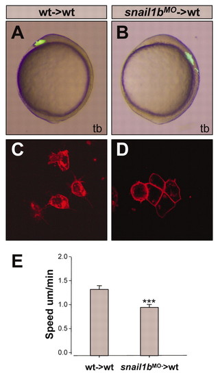
Migratory behaviour of snail1b morphant cells in wild-type hosts. (A,B) Lateral views of wild-type live embryos after transplantation at the shield stage of 10-20 axial mesendodermal cells from wild-type (A) or snail1b morphant (B) fluorescein dextran-labelled embryos. (C,D) Membrane-tagged RFP-labelled grafts from wild-type (C) or snail1b morphant (D) embryos on wild-type hosts at 70% epiboly stage allows the visualisation of differences in individual cell morphology: wild-type cells show protrusions whereas snail1b morphant cells form cohesive clusters and show an epithelial-like morphology. (E) Speed of wild-type and snail1b morphant axial mesendodermal cells in wild-type hosts (P<0.001). In each embryo, the distances of three individual cells were measured (wt→wt, n=7; mo→wt, n=5)
|