
Fig. 4
- ID
- ZDB-FIG-071006-3
- Publication
- Arnaout et al., 2007 - Zebrafish model for human long QT syndrome
- Other Figures
- All Figure Page
- Back to All Figure Page
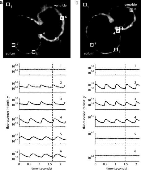
Wild-type embryonic hearts in the Tg(cmlc2:gCaMP)s878 background exhibit atrial and ventricular conduction waves, whereas mutant hearts exhibit atrial, but no ventricular, conduction waves. SPIM videos of live 48 hpf hearts in the Tg(cmlc2:gCaMP)s878 background (see SI Movies 3 and 4) were processed to determine the fluorescence intensity of selected regions of the heart over time. Each selected region has a corresponding number plotted below. The dotted lines mark an arbitrary point in time to facilitate comparison across the different plots. (a) In a wild-type heart, fluorescence intensity varies with time in atrial and ventricular regions of the heart as the wave of depolarization propagates through the heart. This wave represents a wild-type heart rhythm. (b) In a kcnh2s290 mutant heart, fluorescence intensity varies in the atrium, but the ventricle maintains a constant, low-level intensity. The mutant heart lacks a ventricular conduction wave. |
| Fish: | |
|---|---|
| Knockdown Reagent: | |
| Observed In: | |
| Stage: | Long-pec |