
Fig. 3
- ID
- ZDB-FIG-070927-38
- Publication
- Yu et al., 2005 - Semaphorin signaling guides cranial neural crest cell migration in zebrafish
- Other Figures
- All Figure Page
- Back to All Figure Page
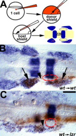
lzr/pbx4 functions non-cell autonomously to control cranial NCC migration. (A) Schematic of the approach for generating cranial NCC mosaics. Donor-derived cells are brown, dlx2 and krox20 expression are in blue. Red dash-lines circle the position of the OV in B-C. (B) In control experiments, wild-type donor cells migrate normally into the second and third cranial NC streams of the pharyngeal arches (arrows) without occupying the cranial NC-free zone (arrowhead) in a wild-type host embryo. (C) In contrast, wild-type donor cells are seen in the normally cranial NC-free zone (arrowhead) after transplantation into a lzr/pbx4 mutant host. Scale bar: 40 μm in B–C. |
Reprinted from Developmental Biology, 280(2), Yu, H.H., and Moens, C.B., Semaphorin signaling guides cranial neural crest cell migration in zebrafish, 373-385, Copyright (2005) with permission from Elsevier. Full text @ Dev. Biol.