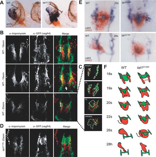
Defective Heart Tube Formation in tal1t21384 Mutant Embryos Despite Normal Fusion of Bilateral Myocardial Precursor Populations (A) Chamber differentiation in wt and tal1t21384 mutant embryos revealed by two-color in situ hybridization showing amhc (atrium) and cmlc2 (atrium and ventricle) at 28 hpf. Chamber differentiation proceeds normal in mutant embryos, but the primary heart tube is malformed with an enlarged atrial inflow region (brackets). (B–D). Two-color immunohistochemistry using anti-tropomyosin (red, myocardium) and anti-gfp (green, vegfr4, endocardium) antibodies. Images were generated as maximum projections of confocal z-stacks (ventral views, anterior to the top). Some yolk platelets show intense autofluorescence at the wavelength used for anti-tropomyosin detection (647 nm). (B) Heart morphogenesis during heart field fusion in wt embryos. The bilateral heart fields fuse dorsal to the endocardium between the 18- and 20-somite stage. Fusion is initiated in the posterior region of the heart. At the 18-somite stage, tropomyosin-positive cells are located ventral to the first aortic arches. (C) Endocardial precursors are ventral to the myocardium in the lateral and posterior regions of the heart. Endocardial and myocardial sheets are closely associated, as relative positions were only revealed after deconvolution of confocal stacks. Four deconvolved images of the confocal image stack in (B) are shown in (C). (D) In tal1t21384 mutant embryos, initial fusion of the myocardial precursor populations occurs normally, although endocardial precursors are absent in the posterior region of the heart field. (E) Primary heart tube formation from myocardial and endocardial precursors in wt and tal1t21384 mutant embryos revealed by two-color in situ hybridization showing cdh5 (endocardium, blue) and cmlc2 (myocardium, red) expression. Dorsal views, anterior is to the top. At the 22-somite stage, wt embryos have formed a cardiac disc, with endocardial cells underlying the circular myocardial primordium. The medial endocardium within the ring of myocardium forms the connection between the endocardium and the aortic arches. In tal1t21384 mutant embryos, anterior closure of the myocardial primordium is defective due to aggregation of endocardial precursors. At the 26-somite stage, wt embryos have formed the primary heart tube and rhythmical contractions begin. In tal1t21384 mutant embryos, heart tube formation is delayed. By this stage, myocardial cells have completed fusion formation at the anterior side of the cardiac disc. (F) Schematic overview of fusion of myocardial precursor regions and heart tube formation in wt and tal1t21384 mutant embryos. Endocardium and aortic arches are in green, myocardium in red.
|

