
Fig. 3
- ID
- ZDB-FIG-070925-55
- Publication
- Albertson et al., 2007 - Fgf8 haploinsufficiency results in distinct craniofacial defects in adult zebrafish
- Other Figures
- All Figure Page
- Back to All Figure Page
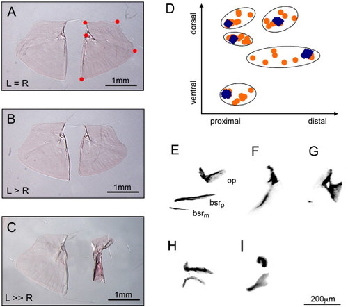
Fig. 3. Aceti282a/+ zebrafish exhibit asymmetry in opercle size and defects in opercle shape. Opercle size was defined as the square root of the sum of squared distances of the five landmarks depicted in panel A and their centoid (e.g., centroid size). Most mutants possessed left and right opercles that were equivalent in size (A). 28%, however, expressed a pronounced asymmetry in opercle size, with the left side typically larger than the right (B, C). Defective opercle shape in aceti282a heterozygous zebrafish mainly affected the width of the opercle (D). Wild-type landmark configurations are depicted as blue diamonds, and aceti282a/+ configurations are orange circles. Landmarks are the same as in panel A. Very little landmark variation was observed in wild-type zebrafish. Heterozygous animals on the other hand exhibited much greater landmark variation, especially along the proximal–distal axis. Opercle defects in aceti282a heterozygotes were distinct from those in aceti282a homozygous recessive mutants (E–I). The wild-type configuration of bones from the hyoid arch is shown in the 7 dpf larvae (E). Elements are arranged along the dorsal–ventral (DV) axis with the medial branchiostegal ray (bsrm) positioned ventrally, the opercle (op) positioned dorsally, and the posterior branchiostegal ray (bsrp) positioned in the middle. Nearly all aceti282a mutants were missing the ventral-most branchiostegal ray (bsrm) and the other two elements were small and misshaped (n = 194). However, a percentage of mutant larvae (∼ 10%) exhibited a range of bone morphologies possibly related to defects in DV patterning of the hyoid arch (F–I); these included fused opercle and branchiostegal rays (F), “opercle-gain” phenotypes (Kimmel et al., 2003) (G), and homeotic transformations of the opercle to a branchiostegal ray (H) and visa versa (I). |
| Fish: | |
|---|---|
| Observed In: | |
| Stage: | Adult |
Reprinted from Developmental Biology, 306(2), Albertson, R.C., and Yelick, P.C., Fgf8 haploinsufficiency results in distinct craniofacial defects in adult zebrafish, 505-515, Copyright (2007) with permission from Elsevier. Full text @ Dev. Biol.