
Fig. 1
- ID
- ZDB-FIG-070925-46
- Publication
- Albertson et al., 2007 - Fgf8 haploinsufficiency results in distinct craniofacial defects in adult zebrafish
- Other Figures
- All Figure Page
- Back to All Figure Page
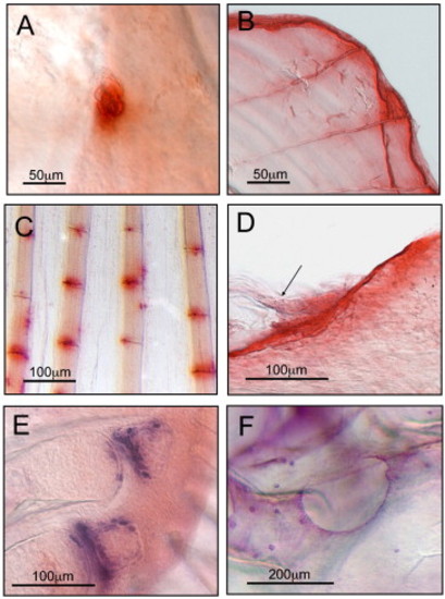
Fig. 1. Images of TRAP (A–D) and AP (E–F) staining in the adult zebrafish. TRAP activity was observed in osteoclasts (A), and in bony structures subjected to continuous growth and remodeling (B–D). An individual osteoclast cell is seen on the surface of the parietal bone (A). TRAP activity was often observed around the distal edges of scales (B). Fin ray joints from all five fins also stained for TRAP, although it was more frequently observed in pectoral and tail fins (pectoral fin, C). Panel D shows TRAP activity on the dorsal surface of the coronoid process of the dentary where the maxillary–mandibular ligament inserts (arrow, anterior is to the left). AP staining of osteoblast cells was observed at sites undergoing endochondral (E) and intramembranous (F) ossification. Endochondral ossification can be seen on pectoral fin radials 4 and 5 (E, dorsal is up). Intramembranous ossification was observed in the lateral line canal on the frontal bone (F, anterior is to the left). |
Reprinted from Developmental Biology, 306(2), Albertson, R.C., and Yelick, P.C., Fgf8 haploinsufficiency results in distinct craniofacial defects in adult zebrafish, 505-515, Copyright (2007) with permission from Elsevier. Full text @ Dev. Biol.