
Fig. 10
- ID
- ZDB-FIG-070920-48
- Publication
- Sharma et al., 2005 - Role of Fyn kinase in signaling associated with epiboly during zebrafish development
- Other Figures
- All Figure Page
- Back to All Figure Page
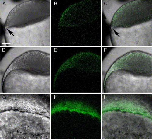
Morphology of the marginal region of the blastodisc in normal zebrafish embryos. Embryos developed from zygotes injected with calcium green-dextran were imaged by confocal microscopy to obtain ‘bright field’ and green fluorescence images demonstrating the distribution of calcium green fluorescence in the embryo. An embryo was imaged at sphere stage (A–C) and at about 20% epiboly (D–I). Images A–F are focused through the equator of the embryo, while in images G–I, the focal plane passed tangentially through the leading edge of the advancing blastoderm. Arrows in panels A and C indicate the position of the YSL. Magnification is indicated by the bar which represents 100 μm. |
Reprinted from Developmental Biology, 285(2), Sharma, D., Holets, L., Zhang, X., and Kinsey, W.H., Role of Fyn kinase in signaling associated with epiboly during zebrafish development, 462-476, Copyright (2005) with permission from Elsevier. Full text @ Dev. Biol.