
Fig. 1
- ID
- ZDB-FIG-070823-38
- Publication
- Xiong et al., 2006 - Tob1 controls dorsal development of zebrafish embryos by antagonizing maternal beta-catenin transcriptional activity
- Other Figures
- All Figure Page
- Back to All Figure Page
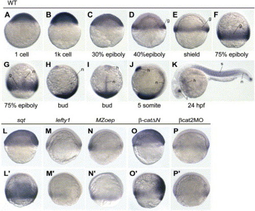
Spatiotemporal Expression Pattern of tob1a and Its Regulation (A–K) Expression pattern of tob1a, detected by whole-mount in situ hybridization, at indicated stages. Embryo orientations: (A–F), (H), and (J), lateral views with the animal pole oriented at the top; (G) and (I), dorsal views with the animal pole oriented at the top; (K), anterior is oriented toward the left. The indicated domains: g, germ ring; h, prechordal/mesoderm/hatching gland; l, lens; n, notochord; s, somites. (L–P′) tob1a expression in embryos injected with indicated mRNA species or in MZoep mutants. Except for (L′), which is a dorsal view, panels are shown in lateral views with the animal pole oriented at the top and dorsal oriented toward the right. (L–P) show the shield stages; (L′–P′) show the 75% epiboly stage. Injection doses: sqt, 0.5 pg; lefty1, 50 pg; β-catΔN, 10 pg; βcat2MO, 20 ng. |
| Gene: | |
|---|---|
| Fish: | |
| Knockdown Reagent: | |
| Anatomical Terms: | |
| Stage Range: | 1-cell to Prim-5 |
| Fish: | |
|---|---|
| Knockdown Reagent: | |
| Observed In: | |
| Stage: | Shield |
Reprinted from Developmental Cell, 11(2), Xiong, B., Rui, Y., Zhang, M., Shi, K., Jia, S., Tian, T., Yin, K., Huang, H., Lin, S., Zhao, X., Chen, Y., Chen, Y.G., Lin, S.C., and Meng, A., Tob1 controls dorsal development of zebrafish embryos by antagonizing maternal beta-catenin transcriptional activity, 225-238, Copyright (2006) with permission from Elsevier. Full text @ Dev. Cell