
Fig. 1
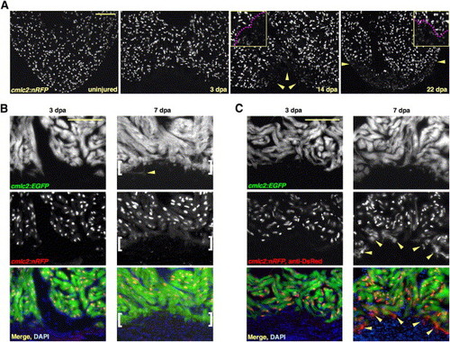
New Myocardium Arises from Undifferentiated Progenitor Cells (A) Sections through uninjured and injured cmlc2:nRFP ventricles. A subpopulation of RFPlo nuclei manifests by 14 dpa (arrowheads), representing ostensibly the entire regenerate by 22 dpa. Magenta line in high-magnification insets delineates RFPhi CMs (above line) from RFPlo CMs. (B) cmlc2:nRFP; cmlc2:EGFP ventricles. EGFP and RFP expression from the cmlc2 promoter is reported at the same basoapical level at 3 dpa. By 7 dpa, an RFPneg front of newly differentiated muscle reporting the faster-fluorescing EGFP (brackets) appears apical to the EGFPposRFPpos portion. Arrowhead indicates an EGFPpos cell process extending into the clot. (C) cmlc2:nRFP; cmlc2:EGFP ventricles stained for DsRed immunoreactivity with an anti-DsRed antibody. At 3 dpa, there is no difference in appearance from the unstained 3 dpa ventricle in (B). By 7 dpa, a front of RFPcyto muscle (arrowheads), representing the most recently differentiated CMs, colabels EGFPpos tissue apical to natural RFPnuc fluorescence. Scale bar = 100 μm. |
Reprinted from Cell, 127(3), Lepilina, A., Coon, A.N., Kikuchi, K., Holdway, J.E., Roberts, R.W., Burns, C.G., and Poss, K.D., A Dynamic Epicardial Injury Response Supports Progenitor Cell Activity during Zebrafish Heart Regeneration, 607-619, Copyright (2006) with permission from Elsevier. Full text @ Cell