
Fig. 5
- ID
- ZDB-FIG-070810-21
- Publication
- Lakowski et al., 2007 - Mechanisms controlling Pax6 isoform expression in the retina have been conserved between teleosts and mammals
- Other Figures
- All Figure Page
- Back to All Figure Page
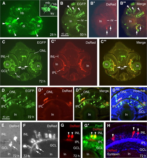
Amacrine cells in the developing zebrafish retina express DsRed. Sections cut through the eye of transiently transgenic embryos at different stages of development. (A) At 28 hpf EGFP, but not DsRed, is expressed by retinal progenitor cells (arrowheads). EGFP-expressing cells are also visible in the diencephalon (di) and hindbrain (*). Inset: lateral view of a 28 hpf embryo showing the plane of section. Rostral is left and dorsal is up. (B) Starting at the time of amacrine cell differentiation (50 hpf), a subset of EGFP-expressing cells begin expressing DsRed (arrows). (C, D) Whereas differentiating cells in the ONL, INL, and GCL expressed EGFP, only amacrine cells appeared to express DsRed. EGFP expression in retinal ganglion cell axons was visible within the optic nerve by 72 hpf (on). Cell nuclei in D are labeled (blue) with Hoechst. (E–H) Sections cut through the eye from four different transiently transgenic embryos that were allowed to develop to 72 hpf. (E, F) DsRed is expressed by displaced amacrines (arrows), which have cell bodies in the GCL. (G) DsRed-expressing cells (red) coexpress Pax6 (immunostained in green). (H) DsRed-expressing cells (red) coexpress syntaxin (immunostained in blue), which is expressed in the dendritic processes and perikarya of all amacrine cells. hb, hindbrain; ln, lens; nr, neural retina. |
Reprinted from Developmental Biology, 307(2), Lakowski, J., Majumder, A., and Lauderdale, J.D., Mechanisms controlling Pax6 isoform expression in the retina have been conserved between teleosts and mammals, 498-520, Copyright (2007) with permission from Elsevier. Full text @ Dev. Biol.