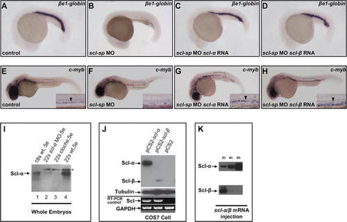
Differences in Protein Expression Levels of Scl-α and -β Confer Their Distinct Functions (A–H) βe1-globin and c-myb expression in scl-sp morphants is rescued by scl-α and -β mRNA. (A–D) show WISH of βe1-globin in 20-hpf control embryos (A), scl-sp morphants (B), scl-sp morphants injected with scl-α mRNA (C), and scl-sp morphants injected with scl-β mRNA (D). (E–H) show WISH of c-myb in 30-hpf control embryos (E), scl-sp morphants (F), scl-sp morphants injected with scl-α mRNA (G), and scl-sp morphants injected with scl-β mRNA (H). Insets in (E–H) are high-magnification (20×) views of the ventral wall of DA, indicated by black arrowhead. In all panels, embryos are in lateral view with anterior to the left. (I) Immunoblotting with Ab-Scl-C antiserum shows the expression levels of Scl-α and Scl-β proteins in wild-type (wt) embryos, clo mutant embryos, and scl-α morphants. Although Scl-β protein was not detectable, immunoblotting of whole embryo protein extracts (50 μg for each sample) showed that Scl-α protein expression increased as embryos developed from the 18-somite (lane 1) to 22-somite (lane 4) stage. The clo mutant embryos (lane 3) and scl-α morphants (lane 2), in which Scl-α protein was not detected, were used as the controls to distinguish the Scl-α protein (arrow) and nonspecific band (asterisk). (J) Immunoblotting with Ab-Scl-C antiserum shows the expression levels of Scl-α and Scl-β proteins in transfected COS7 cells. Western blotting of whole cell protein extracts (10 μg for each sample) prepared from COS7 cells transfected with full-length scl-α, scl-β, or blank vector constructs revealed that the protein level of Scl-α was much higher than that of Scl-β. RT-PCR analysis (bottom) showed similar RNA levels of scl-α and -β in these transfected cells. Tubulin and GAPDH were used as controls for Western blot and PCR, respectively. (K) Immunoblotting with Ab-Scl-C antiserum shows the expression levels of Scl-α and Scl-β proteins in the embryos injected with in vitro synthesized scl-α or -β mRNA. Western blotting of whole embryo protein extracts (50 μg for each sample) from embryos injected with 500 pg of scl-α or -β mRNA showed that, at 3 h post-injection, the Scl-β protein was detected as being at a comparable level to that of Scl-α but dramatically reduced by 4 h post-injection.
|
