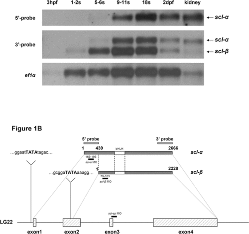
Fig. 1
- ID
- ZDB-FIG-070619-30
- Publication
- Qian et al., 2007 - Distinct Functions for Different scl Isoforms in Zebrafish Primitive and Definitive Hematopoiesis
- Other Figures
- All Figure Page
- Back to All Figure Page
|
Identification of Zebrafish scl-β Isoform (A) Northern blot analysis of scl expression in 3-h, 1- to 2-somite (s), 5- to 6-somite, 9- to 11-somite, 18-somite, and 2-dpf embryos and adult kidney. The 3′-probe (3′ UTR) recognized both scl-α and -β isoforms (middle), whereas the 5′-probe (5′ UTR) detected only the full-length scl-α (top). ef1α was used as the loading control (bottom). (B) Gene structures of scl-α and -β. scl-α consists of 4 exons (boxed regions). scl-β transcript is initiated from the middle region of exon 2, and a potential TATA-box sequence (indicated in bold capital letters) is predicted at position -31 of the transcription initiation site. The black bars indicate the positions of scl-α MO, scl-β MO, and scl-sp MO. The white boxes represent sequences that encode the basic helix-loop-helix domain. |

