
Fig. 7
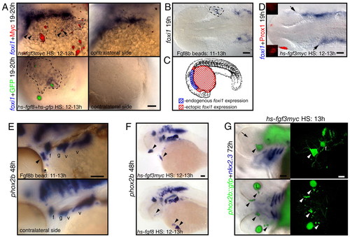
Fgf3 or Fgf8 is sufficient to induce foxi1-positive EB precursors and phox2b-positive EB neurons in wild-type embryos. Zebrafish embryos were injected with hs-fgf3myc or coinjected with hs-fgf8 and hs-gfp plasmids at one-cell stage, heat shocked at 10-13 hpf and then assayed for foxi1 or phox2b expression (blue). Alternatively, embryos were implanted with Fgf8b-coated beads at 11 hpf and assayed for foxi1 and phox2b expression at 19 and 48 hpf, respectively. (A,E,F) Lateral views; (B,D) dorsal views. (A) Ectopic foxi1-positive cells (outlined by dotted line) are immediately adjacent to the Myc-(red) or GFP-positive (green) cells expressing Fgf. Note punctate staining surrounding Myc-positive cells, presumably indicating secreted myc-tagged Fgf3 protein. (B) Ectopic foxi1 expression (dotted line) was induced in the vicinity of Fgf8b beads. (C) Summary of the ectopic Fgf expression experiments. Ectopic foxi1 foci (shown in red) were restricted to the ventral side of the yolk surface, just ventral and anterior to the endogenous foxi1-expression domain. (D) Activation of hs-fgf3myc leads to the anterior expansion of foxi1 domain (arrows) and loss of Prox1 expression in the lens (red). Insets show lateral views of the presumptive lens domain on each side of the embryo. (E) Fgf8b bead (star) induced formation of the ectopic phox2b-positive EB neurons (arrowhead). (F,G) Activation of Fgf3myc or Fgf8 in wild-type (F) or phox2b::egfp transgenic embryos (G) induced ectopic phox2b-positive EB neurons (arrowheads) away from the endogenous phox2b-expression sites. (G) Left panels show overlay of immunofluorescence and brightfield photographs, and right panels show confocal stacks generated from the same embryos. Note ectopic phox2b-positive cells on the ventral surface of the head as well as in the eye (arrowheads). Formation of the ectopic phox2b-positive neurons (green) did not require endoderm pouch tissue, visualized by nkx2.3 (purple). Note complete absence of lens tissue (arrow, |