|
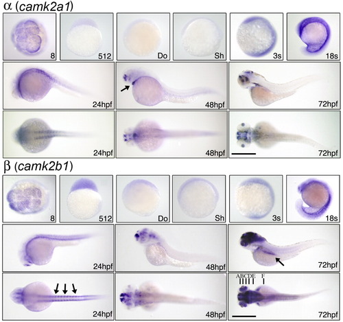
In situ localization of α and β CaMK-II mRNAs. CaMK-II expression was assessed by in situ hybridization with a probe for camk2a1 and camk2b1 at the indicated stages. Stages analyzed in Figures 4-6 include the 8-cell stage (8; 1hpf), the 512-cell stage (512; 2.5hpf), the dome stage (Do; 4hpf), the shield stage (Sh; 6hpf), the 3-somite stage (3s; ~ 11hpf), and the 18-somite stage (18s; ~ 18hpf). The 8-cell stage is an animal poll view; others are lateral except for dorsal views at 24, 48, and 72hpf. Arrow in camk2a1 indicates heart. Arrows in camk2b1 at 24hpf indicate somites and at 72hpf gut. Letters locate cross-sections for Figure 7. Scale bar = 1 mm.
|