
FIGURE
Fig. 4
- ID
- ZDB-FIG-070219-4
- Publication
- Tamme et al., 2002 - The identity and distribution of neural cells expressing the mesodermal determinant spadetail
- Other Figures
- All Figure Page
- Back to All Figure Page
Fig. 4
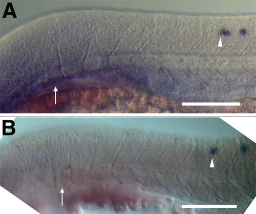
Lateral views of two embryos (A and B) at approximately 24 hpf stained to reveal spt transcription. Dorsal is up and rostral is to the left. DIC microscopy was used to reveal somite boundaries. Consequently, spt-expressing cells in the developing CNS are not seen clearly because they lie in a different focal plane. However, the most rostral cell in each embryo is indicated by a white arrrowhead. The most rostral visible discernible somite is indicated by a white arrow. In both cases there are 6 somites rostral to the most rostral spt-expressing neuron. Scale bars equal 100 μm. |
Expression Data
| Gene: | |
|---|---|
| Fish: | |
| Anatomical Terms: | |
| Stage: | Prim-5 |
Expression Detail
Antibody Labeling
Phenotype Data
Phenotype Detail
Acknowledgments
This image is the copyrighted work of the attributed author or publisher, and
ZFIN has permission only to display this image to its users.
Additional permissions should be obtained from the applicable author or publisher of the image.
Full text @ BMC Dev. Biol.