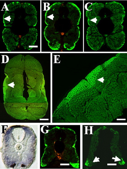
Different levels of green fluorescent protein (GFP) expression in slow and fast muscles of smyd1-gfp transgenic fish. A-C: GFP expression directly photographed on embryonic sections at 4 days postfertilization (dpf) under a confocal microscope. Slow muscles express higher levels of GFP than fast muscles in three transgenic lines of smyd1-gfp transgenic fish. A, line-27; B, line-32; C, line-51. Arrows indicate slow muscles. D,E: Cross-section showing higher levels of GFP expression in slow muscles of line-32 at 2 months old. Slow muscles are indicated by arrows. F: Cross-section shows smyd1 mRNA expression in both slow and fast muscles at 4 dpf, although the staining appeared slightly stronger in superficial slow muscles. G,H: Cross-sections (dorsal on top) showing GFP expressing slow muscles in wild-type transgenic larvae (G) or yot mutant larvae (H) at 4 dpf. Slow muscles are clearly present at the dorsal and ventral myotome in yot mutant embryos. Scale bars = 150 μm in A-C, 500 μm in D, 200 μm in E, 120 μm in G,H.
|
