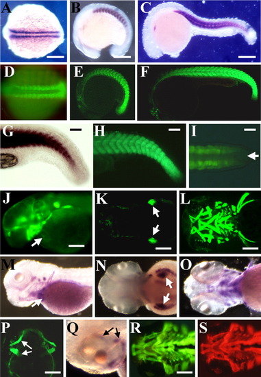
Expression of green fluorescent protein (GFP) reporter gene in developing somites, skeletal, cardiac, head, and fin muscles of transgenic embryos. A-C: In situ hybridization showing expression of smyd1 mRNA in 14 (A), 16 (B), and 22 (C) hours postfertilization (hpf) zebrafish embryos. D-F: GFP expression in developing somites and skeletal muscles of 14 (D), 16 (E), and 22 (F) hpf smyd1-gfp transgenic embryos. G,H: High magnification shows smyd1 mRNA (G) and smyd1-gfp (H) expression in presomitic mesoderm in the tail bud of 24 hpf embryos. I: Dorsal view of GFP expression in presomitic mesoderm but not in the axial mesoderm in the tail bud of smyd1-gfp transgenic embryos at 24 hpf. The arrow indicates axial mesoderm. J: Side view of GFP expression in cardiac muscles of smyd1-gfp transgenic fish at 2 days postfertilization (dpf). K,L: GFP expression in pectoral fin (K, dorsal view) and cranial muscles (L, ventral view) at 2 or 3 dpf, respectively. M-O: In situ hybridization showing expression of smyd1 mRNA in cardiac (M), pectoral fin (N), and cranial muscles (O). Arrows in M and N indicate heart and pectoral fin, respectively. P,Q: Expression of GFP (P, dorsal view) or smyd1 mRNA (Q, side view) in eye muscles of smyd1-gfp transgenic larvae at 5 dpf. R,S: Colocalization of GFP expression and MF20 staining in craniofacial muscles at 4 dpf. GFP expression (R) and MF20 staining (S) were directly observed on the same smyd1-gfp transgenic embryo using a green or a red filter, respectively. Scale bars = 150μm in A-C,R,S, 30μm in G,H, 15 μm in I, 200 μm in M-O,P,Q.
|
