
Fig. 4
- ID
- ZDB-FIG-061110-4
- Publication
- Alt et al., 2006 - Arteries define the position of the thyroid gland during its developmental relocalisation
- Other Figures
- All Figure Page
- Back to All Figure Page
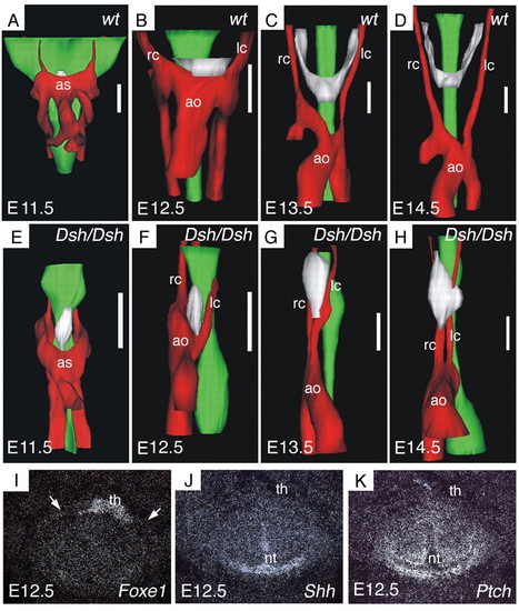
Thyroid morphogenesis is disturbed in short digits (Dsh/Dsh) mutant mice. (A-D) Three-dimensional reconstruction of the cervical region in wild-type mice. (E-H) Same reconstruction of Dsh/Dsh mutant mice. Stages are indicated in the bottom left-hand corner. Red, vascular structures; green, oesophagus; white, thyroid primordium. Scale bar: 140 μm. (I-K) Expression of sonic hedgehog (J) and patched (K) in wild-type E12.5 mouse embryos. Consecutive sections processed for in situ hybridisation, in comparison with the thyroid marker Foxe1 (I). Shh and Ptch are expressed in or around the neural tube, but not in the area of the thyroid. White arrows indicate the position of the carotid arteries (visible in corresponding bright field views). ao, aortic arch; as, aortic sac; lc, left carotid artery; nt, neural tube; rc, right carotid artery; th, thyroid. |