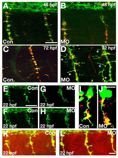
Ventrally projecting motor axons displayed abnormal trajectories in 1.6MO morphants even though CaP axons project normally. (A-D) Motor axon morphology was assayed using SV-2 immunoreactivity (green) in conjunction with a marker of the muscle postsynaptic receptor, α-bungarotoxin (red). At 48 hpf (A,B), little difference was noted in znp-1 immunoreactivity of motor axons of 1.6MO morphants (B) compared with control-injected embryos (A). However, by 72 hpf, there were marked differences (C,D). Motor axons of 1.6MO morphants (D, asterisks) branched more than those of control-injected embryos. Moreover, in morphants (D), there was a reduction in the normal alignment between motor axons and postsynaptic receptors, assessed by the near absence of green and abundance of yellow in C versus the reduction in yellow and increase in green in panel D. (E-L) Injection of 1.6MO did not affect axon outgrowth from CaP when synapses first form. (E-H) At 21-22 hpf, znp-1 immunoreactivity revealed no differences between ventrally projecting CaP axons in either control (E,F) or 1.6MO-injected (G,H) embryos. Embryos were squash-mounted and, consequently, motor nerves on both sides of an embryo were often present in a single confocal section (e.g. F,H). (I,J) CaPs were directly labeled by dye injection (green) at 20 hpf and embryos were fixed at 21-22 hpf and stained for SV-2 immunoreactivity. Injection of either ConMO (I) or 1.6MO (J) did not affect CaP axon morphology. (K,L) In addition to axonal morphology (SV-2, green), postsynaptic specializations, as detected by α-bungarotoxin labeling (red) were not altered by injection of either Con (K) or 1.6 (L) MO. Scale bars: in A, 20 μm for A,B; 30 μm for C; in E, 50 μm for E-H; in J, 10 μm for I,J; in L, 20 μm for K,L.
|
