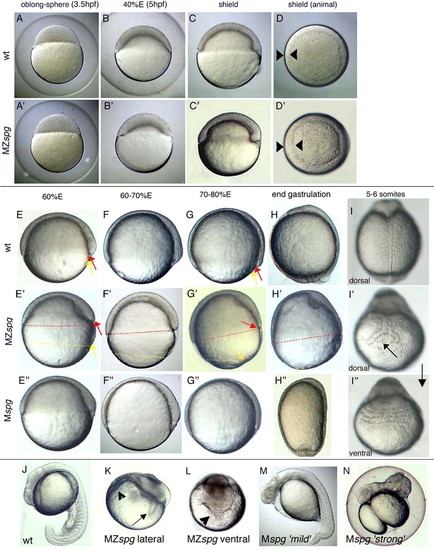
Live morphology of MZspg and Mspg mutant embryos. (A,A') Until sphere stage, mutants are indistinguishable from wild-type embryos. (B,B') Doming and epiboly is inefficient in MZspg embryos and (C,C') the blastoderm fails to flatten. (C-D') The shield forms on time, but the blastoderm has only reached 40% E in MZspg embryos. Shield and germring are thicker when compared with the wild type. (E-H') Epiboly of the YSL and EVL (yellow arrows) is uncoupled from epiboly of the blastoderm (red arrows) in MZspg embryos, which is stalled when the blastoderm covers around 60% of the yolk. (I) The notochord is split in MZspg embryos (I') and the somites fuse on the opposite site (I''). (J-N) After 1 day of development. (K,L) MZspg embryos display severe morphological abnormalities compared with wild type (J) and exhibit massive cell death (arrow in K indicates the split notochord; arrowhead in K and L indicates ventrally fused somites). (E''-H'') Mspg embryos recover completely from their initial epiboly defect until the end of gastrulation. Expressivity of the Mspg phenotype is variable: `strong' Mspg embryos (M) are dorsalized (H'',N) whereas `mild' Mspg embryos are hardly dorsalized.
|
