FIGURE
Fig. 8
- ID
- ZDB-FIG-060628-31
- Publication
- Wei et al., 2004 - The zebrafish Pard3 ortholog is required for separation of the eye fields and retinal lamination
- Other Figures
- All Figure Page
- Back to All Figure Page
Fig. 8
|
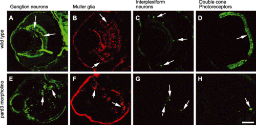
Loss of the Pard3 function did not affect the specification of retinal cells. The retinas of 5 dpf wild-type (A–D) and 5 dpf anti-pard3 morpholino-injected embryos (E–H) were immunostained with zn-8 for ganglion cells (A and E, arrows), anti-carbonic anhydrase for Müller glia (B and F, arrows), anti-tyrosine hydroxylase for interplexiform cells (C and G, arrows), and zpr-1 for double cone photoreceptors (D and H, arrows). Scale bar indicates 50 μm. |
Expression Data
Expression Detail
Antibody Labeling
Phenotype Data
| Fish: | |
|---|---|
| Knockdown Reagent: | |
| Observed In: | |
| Stage: | Day 5 |
Phenotype Detail
Acknowledgments
This image is the copyrighted work of the attributed author or publisher, and
ZFIN has permission only to display this image to its users.
Additional permissions should be obtained from the applicable author or publisher of the image.
Reprinted from Developmental Biology, 269(1), Wei, X., Cheng, Y., Luo, Y., Shi, X., Nelson, S., and Hyde, D.R., The zebrafish Pard3 ortholog is required for separation of the eye fields and retinal lamination, 286-301, Copyright (2004) with permission from Elsevier. Full text @ Dev. Biol.

