Fig. 7
- ID
- ZDB-FIG-060426-4
- Publication
- Reugels et al., 2006 - Asymmetric localization of Numb:EGFP in dividing neuroepithelial cells during neurulation in Danio rerio
- Other Figures
- All Figure Page
- Back to All Figure Page
|
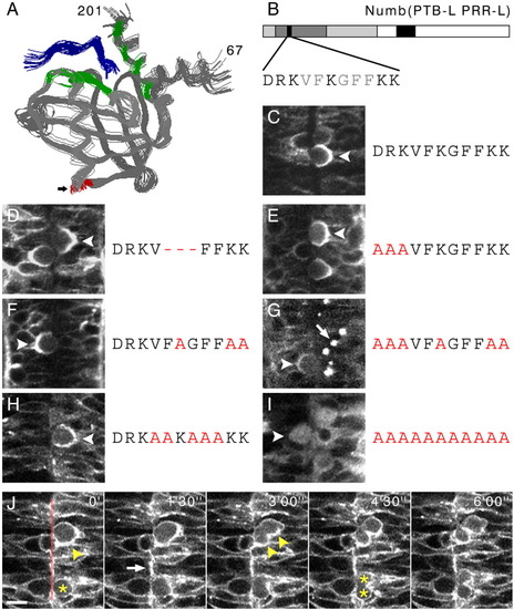
A: Solution structure of the Drosophila Numb PTB domain-Nak peptide complex as described by Zwahlen et al. ([2000]). The PTB domain is coloured in grey, whereas the Nak peptide is coloured in blue. Residues that have direct contacts to target peptides are coloured in green. Alignment of the Drosophila and the zebrafish PTB domain indicates that the additional 11 amino acids of the zebrafish PTBL domain (arrow) would be inserted between amino acids 111 and 112 (highlighted in red). Figure generated using RasMol Vers. 2.7.2.1. B: Sequence of the PTBL insertion. Amino acids with acidic or basic side chains (D, R, K) are coloured in black. Amino acids with nonpolar side chains (V, F, G) are coloured in grey. C: Dorsal view of the neural tube of an embryo injected with numb(PTBL PRRL):egfp mRNA (compare with Fig. 5). D-I: Alanine-scanning mutagenesis of the 11-amino acid insertion. Confocal micrographs of mitotic cells in the neural tube of embryos injected with mRNA made from different mutagenized numb(PTBL PRRL):egfp constructs. See text for details. J: A sequence of five time-lapse frames of two mitotic cells, in the neural tube of embryos injected with numb(DRKAAKAAAKK):egfp mRNA, showing mislocalization of the fusion protein and misorientation of the cell division. The transparent red line marks the midline. Anterior is towards the top. The upper cell (arrowhead in 0′) exhibits a mislocalized Numb(DRKAAKAAAKK):EGFP crescent and subsequently divides with an orientation that is clearly oblique to the plane of the neuroepithelium (arrowheads in 3′00"). The lower cell (asterisks in 0′) divides parallel to the neuroepithelial plane, as is the normal case in neural tube stage (asterisks in 4′30"), but localizes Numb(DRKAAKAAAKK):EGFP ubiquitously around the cell cortex. Note the apical localization of the fusion protein also in the non-dividing cell (arrow in 1′30"). |

