
Fig. 6
- ID
- ZDB-FIG-060426-3
- Publication
- Reugels et al., 2006 - Asymmetric localization of Numb:EGFP in dividing neuroepithelial cells during neurulation in Danio rerio
- Other Figures
- All Figure Page
- Back to All Figure Page
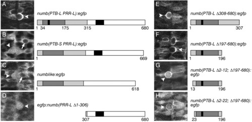
A-H: Comparison of Numb(PTBL PRRL):EGFP, Numb(PTBS PRRL):EGFP, Nbl:EGFP, and five different Numb:EGFP deletion constructs with respect to their localization in dividing cells. Confocal micrographs showing a dorsal view of mitotic cells in the neural tube of embryos injected with the corresponding mRNA. Anterior is towards the top and the midline/apical surface is approximately in the middle of each photograph. See text for details. B-D: Note that the nucleus in non-dividing cells is free of the signal (arrow), whereas in mitotic cells (arrowhead) the signal is found in the entire cell, after the nuclear membrane has broken down. F: Amino acids 1-196 (including the PTBL insertion) are efficiently localized to the basolateral cortex. G: Deletion of amino acids 2-12 impairs cortical and abolishes basolateral localization in dividing cells. H: Impairment of cortical localization is enhanced after deletion of amino acids 2-22. |