
Fig. 4
- ID
- ZDB-FIG-060410-18
- Publication
- Hammond et al., 2006 - The developing lamprey ear closely resembles the zebrafish otic vesicle: otx1 expression can account for all major patterning differences
- Other Figures
- All Figure Page
- Back to All Figure Page
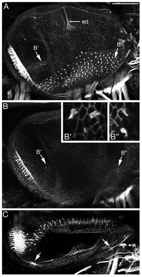
Morphology of the L. fluviatilis otic vesicle at stage 30. (A-C) Projections from confocal z-stacks of a FITC-phalloidin-stained ear showing the actin-rich sensory hair cell stereociliary bundles and general morphology. (A,B) Lateral views, anterior to left, dorsal to top; both are composites of two images. ed, endolymphatic duct. (B) A projection from a lateral subset of the confocal z-stack to show the presumptive cristae clearly, also shown at higher power in insets (B',B''). (C) Dorsal view, anterior to left, medial to top; arrows indicate hair cells in the developing cristae. Scale bars: 50 μm. |