
Fig. 4
- ID
- ZDB-FIG-060216-4
- Publication
- Amacher et al., 1998 - Promoting notochord fate and repressing muscle development in zebrafish axial mesoderm
- Other Figures
- All Figure Page
- Back to All Figure Page
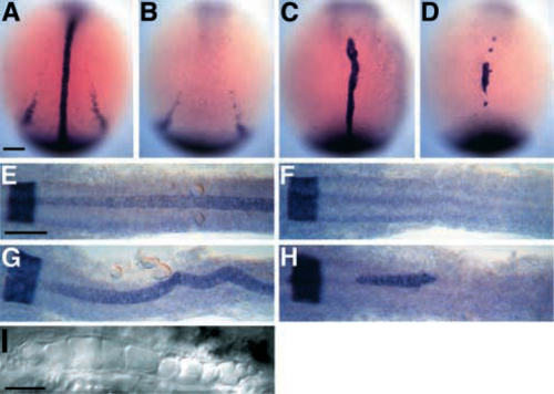
Anterior spt-;flh- midline cells express the essential notochord gene, ntl. (A-D) Dorsal views of ntl expression during early segmentation stages (4-6 somites, 11.3-12 h, anterior to the top). ntl is expressed in wild-type embryos (A) in the developing notochord and in the tailbud (Schulte-Merker et al., 1992, 1994) and, in these overstained embryos, in the more laterally located presumptive pronephric ducts. In flh- embryos (B), midline ntl staining is conspicuously missing. In about half the flh- embryos, there is anterior midline ntl expression in approximately 1-6 cells (not shown). ntl expression in spt- embryos (C) is approximately normal (Hammerschmidt et al., 1996a), except the presumptive pronephros domain is reduced or absent. Midline ntl expression is observed in all spt-;flh- embryos (D), typically in one large anterior patch, and occasionally in a few additional smaller patches, like those shown here. Many spt-;flh- embryos (10/20, 50%) look as shown; however, a significant number (8/20, 40%) have more extensive ntl expression, some of which were recovered from the spt- class after staining. A few spt-;flh- embryos have only about 10 ntlpositive cells (2/20, 10%). The observed number of spt-;flh- embryos was as expected (20/316). (E-H) Dorsal views of krox-20 and ntl staining during later segmentation stages (22 somites, 20 h, anterior to the left) when notochord differentiation is well underway. krox-20 is expressed in a broad band at the extreme left of each photograph that marks a hindbrain segment, rhombomere 5, in all embryos (Oxtoby and Jowett, 1993). In wild-type embryos (E), ntl is expressed in the entire notochord, beginning at the level of rhombomere 4 or 5 and extending posteriorly. Expression is beginning to fade in the anteriormost notochord. No midline ntl expression is observed in any flh- embryos (F). In spt- embryos (G), ntl expression reveals a wide, kinked notochord. Patches of anterior ntl expression are observed in spt-;flh- embryos (H). Most spt-;flh- embryos (8/12) show expression in a patch containing about 20-60 midline cells underlying the posterior half of the hindbrain; three of these embryos have additional smaller clumps located either more anteriorly (as shown in H, see small clump just anterior to rhombomere 5) or more posteriorly at midtrunk levels. A few double mutants (2/12) have two small patches of ntl-expressing midline cells each containing 2-5 cells, and the remaining double mutants (2/12) show no ntl expression at this stage. (I) 30 h Nomarski view of spt-;flh- notochord cells. Scale bars, 100 um (A-H), 50 um (I). |