
Fig. 2
- ID
- ZDB-FIG-060216-2
- Publication
- Amacher et al., 1998 - Promoting notochord fate and repressing muscle development in zebrafish axial mesoderm
- Other Figures
- All Figure Page
- Back to All Figure Page
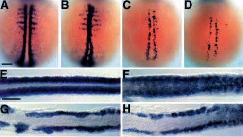
myoD is not expressed in the spt-;flh- midline. (A-D) Dorsal views of myoD expression at early segmentation stages (2-4 somites, 10.7-11.3 h, anterior to the top). In wild-type embryos (A), myoD is expressed in a two bilateral rows of adaxial cells and more laterally in each somite (Weinberg et al., 1996); in flh- embryos (B), myoD is expressed in the midline (Halpern et al., 1995). There are fewer myoD-expressing cells in spt- (C; Weinberg et al., 1996) and spt-;flh- (D) embryos, but they are confined to mesoderm flanking the developing notochord. (E-H) Dorsal views of myoD expression at late segmentation stages (26 somites, 22 h, anterior to the left). In wild-type embryos (E; Weinberg et al, 1996), myoD is expressed in bilateral rows of muscle that flank the notochord. In flh- embryos (F), myoD is expressed in midline mesoderm. myoD expression in spt- (G) and spt-;flh- (H) embryos is strikingly similar, although occasionally a few myoD-positive midline cells are observed in the double mutant tail. Scale bars, 100 um. |