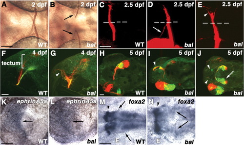
Retinal ganglion cell (RGC) axon defects in bal embryos. K-N are in situ hybridizations. All views are anterior left. A,B: Ventral views of ZN-5-labeled RGC axons in 2 days postfertilization (dpf) wild-type (WT; A) and bal (B) embryos showing ipsilateral projections in bal (arrows in B). C-E: Ventral views of 2.5 dpf WT (C) or bal (D,E) embryos injected with 1,1′-dioctadecyl-3,3,3′ ,3′-tetramethylindocarbocyanine perchlorate (DiI) into eye on bottom. The arrow in D indicates an ipsilateral projection, and the arrowhead in E shows the split optic tract. The dashed line indicates the midline. F,G: Lateral views, contralateral to injected eye, of 4 dpf WT (F) and bal (G) embryos injected with DiI (red) in nasal and DiA (green) in temporal retina. RGC axons extend anteriorly in bal (arrowhead). Injection sites of DiI and 4-4-dihexadecylaminostyryl-N-methylpyridinium iodide (DiA) overlap in G. H-J: Dorsal views of 5 dpf WT (H) and bal (I,J) embryos injected with DiI nasal and DiA temporal. Arrowheads indicate anterior projections, and the arrow denotes ipsilateral tectal projections. K,L: Lateral views of 48 hpf WT (K) and bal (L) embryos showing ephrinA5a in the retina (arrow). M,N: Ventral views of 48 hpf WT (M) and bal (N) embryos showing foxa2 along optic pathway (arrowheads) and in midbrain (arrows). E denotes eye. Scale bars = 60 μm.
|
