
Fig. 6
- ID
- ZDB-FIG-060112-23
- Publication
- Paulus et al., 2006 - Zebrafish bashful/laminin-alpha1 mutants exhibit multiple axon guidance defects
- Other Figures
- All Figure Page
- Back to All Figure Page
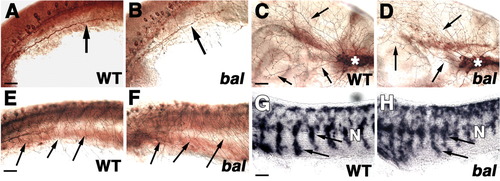
Other peripheral axons and migrating neural crest cells are normal in bal. A-F: Neurons were labeled with anti-α-tubulin (A,B), or ZN-12 (C-F). All are lateral views with anterior to the left. A,B: The 24 hours postfertilization (hpf) wild-type (WT; A) and bal (B) trunks showing posterior lateral line ganglion (PLLg) axons at same somite level (arrows). C,D: The 24 hpf WT (C) and bal (D) heads showing trigeminal axons extending from the trigeminal nucleus (asterisk) along the surface of the head (arrows). E,F: The 24 hpf WT (E) and bal (F) trunks showing Rohon-Beard axons extending along the surface of the trunk (arrows). G,H: In situ hybridization with crestin in 25 hpf WT (G) and bal (H) trunks showing ventral streams of neural crest cells (arrows). N denotes notochord. Scale bars = 40 μm. |