Fig. 2
- ID
- ZDB-FIG-051108-1
- Publication
- Walshe et al., 2003 - Fgf signalling is required for formation of cartilage in the head
- Other Figures
- All Figure Page
- Back to All Figure Page
|
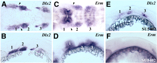
Migrating neural crest cells are recipients of Fgf signals. (A, B) Dorsal (A) and lateral (B) views of 24-hpf embryos showing expression of Dlx2 to show the locations of the three pharyngeal neural crest streams (numbered). (C, D) Dorsal (C) and lateral (D) views of 24-hpf embryos showing expression of Erm in the second and third neural crest streams but little expression in the first stream that migrates adjacent to the isthmus. Transcripts are also detected in the neural tube associated with the isthmus (i) and are detected more weakly in rhombomere boundaries in (C). (E, F) Treatment of embryos with 100 μM SU5402 between 18 and 24 hpf: Dlx2 expression shows that all three neural crest streams are still present (E) but Erm is undetectable (F). Arrowheads in (A) and (C) indicate the position of the first pharyngeal pouch. |
| Genes: | |
|---|---|
| Fish: | |
| Condition: | |
| Anatomical Terms: | |
| Stage: | Prim-5 |
Reprinted from Developmental Biology, 264(2), Walshe, J. and Mason, I., Fgf signalling is required for formation of cartilage in the head, 522-536, Copyright (2003) with permission from Elsevier. Full text @ Dev. Biol.

