
Fig. 1
- ID
- ZDB-FIG-050817-1
- Publication
- Sonawane et al., 2005 - Zebrafish penner/lethal giant larvae 2 functions in hemidesmosome formation, maintenance of cellular morphology and growth regulation in the developing basal epidermis
- Other Figures
- All Figure Page
- Back to All Figure Page
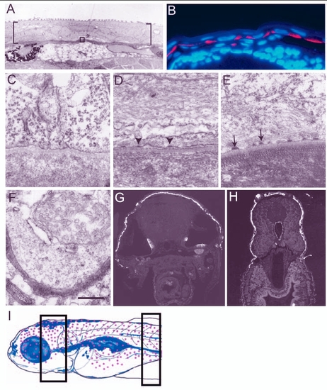
Establishment and maturation of basal domain of the basal epidermal cells during larval development. (A,C-F) Electron micrographs of epidermis. (B,G,H) Histological sections of 3- to 5-day-old larvae stained for keratin using Ks pan1-8 antibody (red in B and white in G,H; blue in B is the nuclear stain DAPI). (A) Larval epidermis, marked by brackets, is bilayered. Asterisk indicates the basal epidermal cell and the area equivalent to that included in the box was analysed from 3- to 5.5-day-old larvae to assess the formation of hemidesmosomes in C-E. (B) Keratin is localised basally in the basal epidermis of 3-day-old larvae. (C-E) Hemidesmosomes are not present in 3-day-old larvae (C); however, premature hemidesmosomes (arrowheads in D) and mature hemidesmosomes (arrows in E) are present in 4.5- and 5.5-day-old larva, respectively. (E,F) Although hemidesmosomes are present in the dorsal epidermis (E), they are absent in the ventral epidermis covering the lower jaw of 5.5-day-old larvae (F). Keratin is also absent in the ventral or ventrolateral epidermal cells of 5-day-old larvae (G,H). Schematic drawing (I) of 5.5-day-old larvae (based on whole-mount keratin staining, immunohistology and electron microscopy data), indicating the epidermal domain (red dots) marked by keratin expression and presence of hemidesmosomes. Sections analysed by electron microscopy were made from the body region marked with rectangles in I. Scale bar: 8.9 µm in A; 40 µm in B; 543 nm in C-F. |