
Fig. 4
- ID
- ZDB-FIG-050721-4
- Publication
- Schweitzer et al., 2005 - Expression of collapsin response mediator proteins in the nervous system of embryonic zebrafish
- Other Figures
- All Figure Page
- Back to All Figure Page
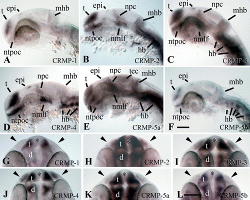
Expression of CRMP mRNAs in the head of 24 hpf embryos. Side views (A–F) or top views (G–L) of whole-mounted embryos with the yolk sac removed are shown. Probes used are indicated in the individual panels. (A–F) (rostral is left, dorsal is up) CNS regions showing expression of the CRMP mRNAs are indicated in the photomicrographs, with the exception of the midbrain–hindbrain boundary (mhb) that is always indicated for orientation, but shows a strong signal only for CRMP-2, -3 and -5a (tec=tectum mesencephali; all other abbreviations as in Fig. 3). (G–L) (rostral is up) Olfactory placodes (always indicated by arrowheads) express CRMP-2, -3, -4 and -5a mRNAs. Telencephalon (t) and diencephalon (d) express all CRMP mRNAs, but not in the proliferating ventricular zone (asterisks). The eyes, which are already lightly pigmented at 24 hpf, are negative for all CRMP mRNAs. Scale bar in F=100 μm for A–F; scale bar in L=100 mm for G–L. |
| Genes: | |
|---|---|
| Fish: | |
| Anatomical Terms: | |
| Stage: | Prim-5 |
Reprinted from Gene expression patterns : GEP, 5(6), Schweitzer, J., Becker, C.G., Schachner, M., and Becker, T., Expression of collapsin response mediator proteins in the nervous system of embryonic zebrafish, 809-816, Copyright (2005) with permission from Elsevier. Full text @ Gene Expr. Patterns