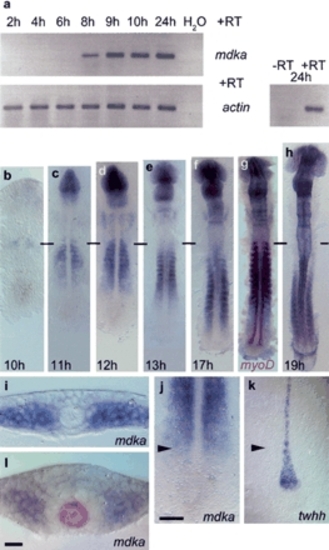
Mdka expression during zebrafish embryogenesis. (a) RT-PCR analysis: mdka transcription is first detected at 8 hpf (h); actin expression was used for calibration. -RT control using actin primers without addition of reverse transcriptase. (b-h) RNA in situ hybridization on 10-19-h embryos showing advancing mdka transcription in the paraxial mesoderm; horizontal bars mark the position of the first somite. (g) Double staining with myoD at 17 h shows progressive loss of mdka transcription in anterior somites. (i) mdka is expressed at the six-somite (6s; 12 hpf) stage in cells of the unsegmented paraxial mesoderm when convergence of the neural plate has just started. (j,k) The front of advancing mdka expression (j) demarcates the position of twhh restriction in MFP precursors (k) as indicated by arrowheads that also show level of transverse section in i. (l) At later stages, mdka is expressed in paraxial mesoderm and also the anterior neural keel, but excluded from a ring of cells surrounding the shh-positive notochord and MFP (8s stage, shh labeled in red). In dorsal views, anterior is to the top in b-h,j,k. Bars: j, 10 µm; l, 50 µm.
|
