- Title
-
The development of vision in the zebrafish (Danio rerio)
- Authors
- Easter, S.S., Jr. and Nicola, G.N.
- Source
- Full text @ Dev. Biol.
|
Photomicrograph of a dorsal view of a fish viewed in the dissecting microscope. The lines indicate the body axis and the planes of the two pupils. The clockwise (CW) and counterclockwise (CCW) directions are indicated. |
|
Retinal image at the appropriate plane. Arrows indicate the edge of the image of the substage condenser aperture diaphragm (SCAD). (a–c) All three panels are micrographs of eyes, focused at the plane of the photoreceptor/pigmented epithelium interface. (a, b) These illustrate the same eye, from a 108 hpf fish, surgically isolated and positioned with the pupil looking into the condenser beam, away from the objective. The image of the SCAD is shown, pinched down in a and opened wide in b. (c) This is a dorsomedial view of a right eye in a live 85 hpf fish, lateral up and to the right, anterior up and to the left. The image of a grating inside the SCAD appears on the dorsal retina. The cone layer is evidenced in the granular texture, most visible in the bright stripes. (d, e) These are two views of the same field, at different planes of focus. (d) A freshly isolated lens (L) adheres to a fragment of the retina (R). The plane of focus is at the level of the lens center. (e) The plane of focus is at the back focal plane of the lens, showing a sharp image of a grating (finer than the one in c) inside a SCAD. (f) A dark-reared 128 hpf fish, showing that the image of the grating and the SCAD are in focus at the level of the cones, whose ellipsoids are visible (arrowheads). The inset shows a less magnified view of this same field. Scale bar in e applies to a, b, d, and e, and the one in f applies to c and f. |
|
Image at an inappropriate plane. (a) The upper sketch shows the setup; a larval zebrafish was positioned obliquely on a microscope slide, inside a water-filled chamber (not shown), with one eye oriented downward toward the condenser lens. The lower sketch shows the larva in more detail, with the parallel rays from the SCAD below indicated by the vertical arrows. The image of the SCAD is formed by the ocular lens at the location given by the convergence of the dashed lines. The letters b, c, and d indicate the planes at which the pictures in b, c, and d were focused (d, the lens center; c, the photoreceptor/pigmented epithelium interface; and b, the plane of the image of the grating). (b–d) These are photomicrographs of the same 68 hpf larva, anterior to the left, viewed dorsolaterally with the right eye (RE, toward the top) facing down and the left eye (LE) facing up. The yolk sac (YS) and ear (E) are indicated. The pictures were taken at the three different focal planes shown in a. The image (unlabeled arrow) lay behind the right retina, about twice as far from the lens center as the photoreceptor/pigmented epithelium interface, so the eye was hyperopic (far-sighted). The scale bar applies to b, c, and d. |
|
Development of the lens. All panels show a dorsoventral section through the lens, dorsal up, lateral to the right. (a) 24 hpf. The lens (L) is spherical but still attached to the skin and surrounded on three sides by the retina (R). (b) 30 hpf. The cornea (C) is now separate, and the cuboidal epithelium (CE) on the lateral surface is distinct from the less structured core (*). (c) 36 hpf. The onionskin organization of the core is now apparent. (d) 48 hpf. The core is noticeably denser than the outside of the lens, and the cuboidal lateral epithelium contrasts with the concentrically organized medial side. (e) 60 hpf. The dense core has enlarged and the concentric laminae are thinner. The medial side begins to bulge, giving the lens an egg shape. (f) 72 hpf. Most of the surface of the lens now touches the retina, the egg shape remains, and the dense core is larger and denser. The scale bar in f applies to all panels. |
|
Ocular morphology and retinal magnification factor. An optical horizontal section of the eye of a live 72 hpf zebrafish, anterior to the left, medial down. The differential interference contrast reveals the retinal inner plexiform layer (IPL). The lens (L) and the inner surface of the retina are in contact. The lines all originate at the center of the lens, the optical center of this eye. The upper two are connected to the peripheral edges of the retina, and the angle subtended by the retina is the retinal field. The lower lines are 125 μm long (lens radius x 2.5), the predicted focal length of the lens, and extend to the vicinity of the photoreceptor/pigmented epithelium interface. |
|
Retinal development between 72 and 96 hpf. The left column shows sections from 72 hpf fish, the right from 96 hpf fish. Each horizontal pair of photos (a and b, c and d, etc.) have the same magnification, given by the scale bar on the right. (a, b) Transverse sections through the heads passing through the centers of the eyes. Dorsal is up. The brain (B), mouth (M), retina (R), and lens (L) are indicated. The boxes labeled e and f indicate the regions shown in e and f. (c, d) More highly magnified views of the eyes in the preceding micrographs. The various layers are indicated: pigmented epithelium (PE), outer nuclear layer (ONL), outer plexiform layer (OPL), inner nuclear layer (INL), inner plexiform layer (IPL), ganglion cell layer (GCL), and optic fiber layer (OFL). The older eye is larger and slightly more developed. The dark lines indicate the boundaries of the functional retina, the sector containing both plexiform layers and photoreceptor outer segments. Lateral to the functional retina lie the lens, the iris (I), and the retinal germinal zone (GZ). (e, f) More highly magnified views (see boxes in a and b) showing the outer segments (brackets) and the medial rectus muscle (MR) enveloped by dashed lines. In the older fish, the outer segments are longer and the medial rectus is larger and has more myofibrils (e.g., curved arrows). (g, h) Tangential sections through the outer nuclear layer of the nasal retina, showing the transversely cut cone nuclei (Nu) in the central field of both panels and the inner segments (IS) in the surrounding area. |
|
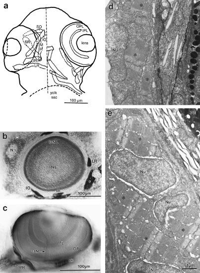
Extraocular muscles. (a) Camera lucida drawing of ventral view of the head of 96 hpf fish which had been reacted against the ZM-1 antibody. To the right of the midline (dashed line), the eye with its lens and two plexiform layers is indicated, as are the head muscles not associated with the eye. To the left, the six extraocular muscles are shown, with the convention that more ventral structures are outlined in solid, and masked outlines (of eye or muscles) are dashed. The six muscles are the superior oblique (SO), the inferior oblique (IO), the medial rectus (MR), the superior rectus (SR), the inferior rectus (IR), and the lateral rectus (LR). (b) Parasagittal (equatorial) section of a 96 hpf eye, dorsal up, rostral left, with approximately transverse sections of five of the six extraocular muscles. Only the medial rectus is missing, because it inserts more medially than the others. The nose (N) is indicated, as are the muscles not associated with the eye (*). (c) Optical section in the horizontal plane of a whole-mounted 96 hpf fish, lateral up, rostral left, showing the belly of the inferior rectus and the insertion of the medial rectus. The plexiform layers and the intraretinal optic nerve (ON) are also shown. (d, e) Electron micrographs of oblique sections through extraocular muscles in 72 and 96 hpf fish, respectively. The scale bar in e applies to both. The melanin in the pigmented epithelium (PE) and the reflective plates in the sclera (SC) provide landmarks for the edge of the eye. The nuclei (Nu) and myofibrils (*) are indicated. The older muscle is larger and more fully packed with myofibrils than the younger. |
Reprinted from Developmental Biology, 180, Easter, S.S., Jr. and Nicola, G.N., The development of vision in the zebrafish (Danio rerio), 646-663, Copyright (1996) with permission from Elsevier. Full text @ Dev. Biol.


