
- Title
-
Evolution of the fish heart by sub/neofunctionalization of an elastin gene
- Authors
- Moriyama, Y., Ito, F., Takeda, H., Yano, T., Okabe, M., Kuraku, S., Keeley, F.W., Koshiba-Takeuchi, K.
- Source
- Full text @ Nat. Commun.
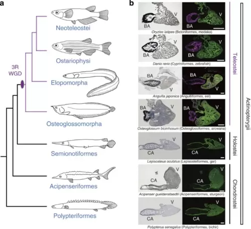
Anatomy and histology of OFTs in actinopterygians. (a) Phylogeny of actinopterygian species. (b) Anatomy and histology of OFTs in representative actinopterygian species. Left panels are results of Elastica van Gieson staining, which visualizes the accumulation of elastic fibres. Note that the elastic fibres of BAs in teleosts are abundant, while those of CAs in non-teleost fishes are restricted to the inner lining. Right panels are results of double immunohistochemistry against α-sarcomeric actinin (cardiac muscle, green) and myosin light-chain kinase (smooth muscle, magenta). Note that teleost BAs are composed of smooth muscle, while CAs in non-teleost fish are composed of cardiac muscle. The phylogenetic timing of acquisition of BA is coincident with 3R WGD. CA, conus arteriosus; V, ventricle. Scale bars, 400 μm. |

Molecular phylogeny and expression patterns of elna and elnb in teleost.(a) Molecular phylogenetic tree of elastin genes (elna/elnb) inferred using 83 amino-acid sites (shape parameter of the gamma distribution alpha=0.67). The gene duplication between elna and elnb is indicated with the black diamond. (b) Syntenic relationship of elna/elnb and limk1a/1b. (c) Molecular phylogenetic tree of limk1 genes inferred using 303 amino-acid sites (alpha=1.03). The gene duplication between limk1a and limk1b is indicated with the black diamond, while a more ancient gene duplication is shown with the grey diamond. The limk1a-1b duplication is shown to have occurred early in the teleost fish lineage, coinciding with 3R WGD. (d–i) Expression patterns of elna and elnb in zebrafish embryos at 3 dpf (d,e,g,h) and 4 dpf (f,i). Arrowheads indicate signals in the BA. Scale bars, 200 μm. (j–m) Expression patterns of elna and elnb in medaka (j,l) and stickleback (k,m) embryos. Note that elnb is expressed only in the BA in both medaka and stickleback embryos. Scale bars, 200 μm. (n–q) Double immunohistological staining of developing OFTs in zebrafish (n,o) and Polypterus (p,q) embryos against α-sarcomeric actinin (cardiac muscle, green) and myosin light-chain kinase (smooth muscle, magenta). Smooth muscle cells appear in the zebrafish BA at 4 dpf, but could not be detected in the Polypterus CA. A, atrium. Scale bars, 50 μm. EXPRESSION / LABELING:
|
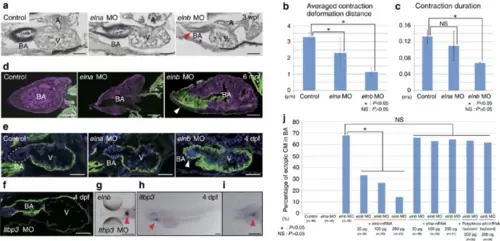
Functions of elna and elnb in BA development. (a) Morphology of BA and elastin accumulation in elna and elnb zebrafish morphants at 3 weeks post-fertilization (wpf). Elastica van Gieson staining highlights hypoplasia and decreased elastin accumulation in elnb morphant BA (arrowhead). Scale bars, 50 μm. (b) Averaged contraction deformation distances in control, elna and elnb morphant BAs. Averaged contraction deformation distances in the elna and elnb BA are significantly reduced compared with that of the control. n=4 each. *P<0.05, NS: P>0.05 (c) Contraction durations in control, elna and elnb morphant BAs. Contraction duration in elnb BA is greatly reduced compared with that of the control. n=4 each. *P<0.05, NS: P>0.05 (d) Anatomy and histology of BA in control, elna and elnb morphants at 6 months post-fertilization (mpf) using double immunohistochemistry; α-sarcomeric actinin (cardiac muscle, green) and myosin light-chain kinase (smooth muscle, magenta). Ectopic cardiomyocytes are observed in elnb morphant BA (arrowhead). Scale bars, 200 μm. This is also the case with embryonic hearts (e). Scale bars, 50 μm. (f) Anatomy and histology of BA in ltbp3 morphants at 4 dpf. Ectopic cardiomyocytes are observed in ltbp3 morphant BA with deformation of heart morphology. Scale bar, 50 μm. (g) elnb expression pattern in ltbp3 morphants at 4 dpf. Expression pattern of elnb is not altered in ltbp3 morphants. Arrowhead indicates elnb expression. Scale bar, 200 μm. (h,i) ltbp3 expression pattern at 4 dpf embryos. ltbp3 is expressed in the BA (arrowheads). Scale bars, 200 μm. (j) Functional divergence of elna and elnb in morphogenesis of the BA. Rescue experiment of elnb morphants by injection of elna, elnb and Polypterus eln full-length mRNAs. Percentages of ectopic cardiomyocytes in the BA are shown in each condition. elna and Polypterus eln mRNA did not rescue the elnb morphant phenotypes while elnb mRNA did. *P<0.05, NS: P>0.05. Dashed lines depict outlines of the BA. Values are reported as mean±s.d. and P values were determined by the t-test. NS, not significant. |

Cell fate determination is altered in elnb morphant BA. (a,b) Fate map of BA (blue) and cardiogenic (red) progenitors in anterior lateral plate mesoderm according to Hami et al.45 Scale bars, 200 μm. (c,d) Example of fate mapping result by injection of fluorescent tracer DiI. Arrowhead indicates injected DiI. Scale bars, 50 μm. (e) Result of cell lineage tracing experiment. In all cases, BA progenitor field to BA or cardiogenic and cardiogenic progenitor field to BA or cardiogenic in both elna and elnb morphants, no statistically significant differences were observd. NS: P>0.05. (f,g) Lengths of BA and ventricle in control, elna and elnb morphants. In all cases no statistically significant differences were observed. NS: P>0.05. (h) Anatomy and histology in yap morphant hearts at 4 dpf. Ecotopic cardiomyocytes are observed in BA (arrowhead). Scale bar, 50 μm. (i) Percentage of ectopic cardiomyocytes in the BA of elnb morphants (control) and yap morphants. (j) elnb expression in yap morphants. elnb expression is not changed in yap morphants (arrowhead). Scale bar, 200 μm. Values are reported as mean±s.d. and P values were determined by the t-test. |

3R WGD and subsequent sub/neofunctionalization of an elastin gene contributed to acquisition of BA in teleost evolution (a) Subfunctionalization and neofunctionalization of elastin genes in actinopterygian evolution. The 3R WGD duplicated elastin into elna and elnb and subfunctionalization occurred between these two genes. After duplication of these genes, mutations were accumulated in the gene body of elnb, and elnb acquired a new function for BA morphogenesis; neofunctionalization occurred. (b) Morphogenesis of BA by elnb in zebrafish heart development. Regionalization of heart such as A, V and OFT is completed by 2 dpf. At 3 dpf, elnb is expressed specifically in OFT. Cardiac progenitor cells in OFT differentiate into smooth muscle cells and elastic and smooth muscle-composed BA is generated. |