FIGURE SUMMARY
Title

lmo4a Contributes to Zebrafish Inner Ear and Vestibular Development via Regulation of the Bmp Pathway

Authors
Sun, L., Ping, L., Gao, R., Zhang, B., Chen, X.
Source
Full text @ Genes (Basel)

Spatiotemporal expression of lmo4a during the first 2 days post-fertilization. The spatiotemporal expression of lmo4a was examined by using both STOmics DB (AH) and in situ hybridization (IN). (AE) Single-cell RNA sequencing data showed concentrated expression of lmo4a in otic placode and otic vesicle at 18 and 24 hpf. (FH) Stereo sequencing showed concentrated expression of lmo4a in otic vesicle at 18 and 24 hpf. (I,J) Maternal lmo4a expression is visible at the ovum stage and 1 hpf. (K) Gastrula period showing lmo4a expression. (L,M) Lateral view at 18 hpf shows restricted expression in the somatic mesoderm and otic vesicle. (N,O) Lateral view at 48 hpf shows lmo4a concentrated expression in the hindbrain and otic vesicle.

Morpholino-targeted knockdown and targeted knockout of lmo4a. (A) Bands of 483 bp and 324 bp represent correctly spliced and artificially mis-spliced lmo4a mRNA. Colored boxes indicate exons. Arrows signify primers used for RT-PCR and the black bar depicts lmo4a MO. (B) Protein alignment including the truncated protein generated by lmo4a MO. (C) RT-PCR of control embryos and those injected with 2 pg lmo4a MO showing effective but incomplete disruption of splicing (m, 100 bp ladder marker). (DG) PCR and Sequencing analysis showing the lmo4a knockout efficiencies of the four gRNA targets.

Detailed phenotypic and behavioral changes in lmo4a morphants. All panels represent lateral views of ears. (AD) Otic morphology of lmo4a morphants at different stages displaying smaller otocysts and thicker otic epithelia compared to WT. (E,G) Hair cell number was reduced in lmo4a morphants, as determined from triple labeling with phalloidin (red, staining the cell framework and stereocilium), anti-myo6 (green, uniformly distributed throughout hair cell cytoplasm) and DAPI (blue, labeling the cell nucleus). (F,H) Embryos were double-labeled with phalloidin and anti-HuC (green, staining SAG), which revealed an evident reduction in the area of SAG in lmo4a morphants. (I,J) Statistical analysis of hair cell numbers of utricle and saccules between control and lmo4a morphants showed significant differences (*** p < 0.001, n = 20). (K) Statistical analysis of the area of SAG between control and lmo4a morphants showed significant differences (*** p < 0.001, n = 20). (L) Anterior, posterior and lateral protrusions were observed in control larvae at 48 hpf; (M) Anterior, posterior and lateral protrusions were fused while ventral protrusions grew out in the control group at 55 hpf. (N) The hubs of semicircular canals exhibited regular shapes in the control group at 72 hpf. (O) No protrusions were visible in lmo4a morphants at 48 hpf. (P) Only lateral protrusions were visible in lmo4a morphants at 55 hpf. (Q) Only lateral protrusions were visible in lmo4a morphants at 72 hpf.

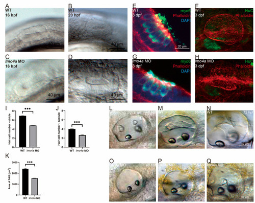
Phenotypic replication and behavioral tests of lmo4a KO embryos. All panels of photos represent lateral views of ears. (AH) Otic morphology of lmo4a KO embryos at different stages displaying smaller otocysts and thicker otic epithelia compared to WT. (I) Statistical analysis confirmed that the otocyst size of lmo4a KO group was significantly smaller at 31, 48, 72 hpf and 15 dpf (*** p < 0.001, n = 20). (J) Swimming paths of WT and lmo4a KO zebrafish were tracked, showing a more active pattern in the KO group. (K,L) The frequencies of both clockwise rotation and counterclockwise rotation were significantly higher in the KO group (* p < 0.05, n = 4).

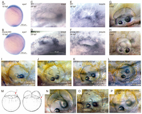
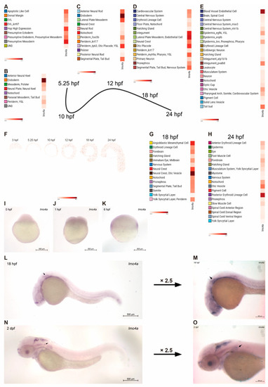
Expression patterns of marker genes of PPE and bmps in lmo4a morphants and rescue of deficiencies in lmo4a morphants via blockage of Bmp signaling. All panels represent lateral views. (A) Expressions of eya1 in WT embryos at 10 hpf. (B) Expressions of eya1 in lmo4a MO embryos were decreased. (C) Bmp4 was expressed strongly at the anterior and posterior ends but weakly in the lateral and dorsal regions of the vesicle in the control group at 28 hpf. (D) Ectopic expression of bmp4 was observed in the dorsal region, with extended original anterior and posterior regions of expression in lmo4a morphants at 28 hpf. (E) bmp2b was expressed strongly at the anterior, posterior and ventral regions in the control group at 36 hpf. (F) Dispersed distribution of bmp2b throughout the vesicle in lmo4a morphants at 36 hpf. (G,H) Chordin (chd) morpholino could replicate the lmo4a MO phenotype with immature protrusions to an extent. (I,J) Heat shock of tBR at 22 hpf successfully rescued the semicircular canal phenotype of lmo4a MO. (K,L) DM knockdown of Bmp at 22 hpf achieved rescue, but to a lower extent. (MP) Rescue was achieved by injecting lmo4a mRNA at the two-cell stage in lmo4a morphants.

Potential molecular mechanisms linking Bmps and lmo4a. (A) Lmo4 acts downstream of the Bmp signaling pathway. (B) Lmo4 acts as a regulator of Bmps. (C) Lmo4 and Bmps function in two parallel pathways with mutual regulatory interactions.

Acknowledgments
This image is the copyrighted work of the attributed author or publisher, and ZFIN has permission only to display this image to its users. Additional permissions should be obtained from the applicable author or publisher of the image. Full text @ Genes (Basel)