
- Title
-
Expression of olfactory receptor genes in non-olfactory tissues in the developing and adult zebrafish
- Authors
- Jundi, D., Coutanceau, J.P., Bullier, E., Imarraine, S., Fajloun, Z., Hong, E.
- Source
- Full text @ Sci. Rep.
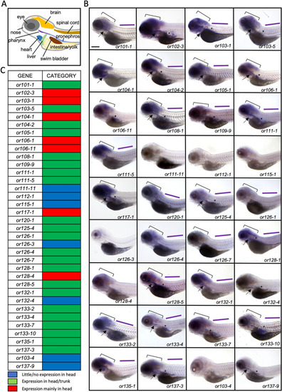
Olfactory receptor genes are expressed throughout the body of zebrafish larvae. (A) Schematic of a lateral view of zebrafish larva illustrating different organs. (B) Lateral views of 6-day-old larvae processed by whole mount in-situ hybridization showing expression of 36 olfactory receptor genes throughout the head and body. Panels are ordered in numerical order of the gene names, except for or103-4 and or137-9, which were not found in the RNA-seq data and placed at the end. The head (bracket), pharynx (arrow), pancreas (asterisk) and trunk (purple line) are indicated. Scale bar = 200 μm. (C) Summary of olfactory receptor gene expression classified into categories (1) little/no expression in the head (blue), (2) expression in the head and trunk (green), (3) strong expression in the head (red). EXPRESSION / LABELING:
|
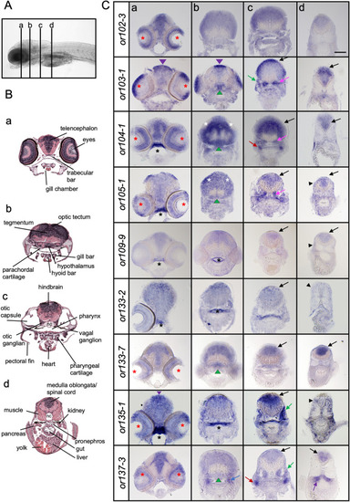
Olfactory receptor genes are expressed in distinct areas in the larval head. (A) Lateral view of a zebrafish larva showing the anterior–posterior levels (a–d) of the transverse sections in (B) and (C). (B) Images of transverse sections of 7-day-old larvae with different structures annotated. Sections represent the forebrain (a), midbrain (b), hindbrain (c) and posterior hindbrain (d). Modified from Bio-Atlas (Jake Gittlen Laboratories for Cancer Research). (C) Transverse sections of 6–7-day-old zebrafish larvae processed by in situ hybridization showing expression of olfactory receptor genes in different areas of the head. The stainings are localized in the dorsal midline (purple arrowhead), eyes (red asterisk), roof of pharynx (black asterisk), optic tectum (white asterisk), hypothalamus (green arrowhead), otic capsule (green arrow), otic ganglion (pink arrow), anterior lateral line ganglion (blue arrow), pronephros (purple arrow), vagal ganglion (red arrow), hindbrain (black arrow) and muscle (black arrowhead). Scale bar = 100 μm. EXPRESSION / LABELING:
|
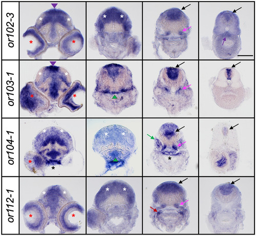
Spatial regulation of olfactory receptor gene expression during development. Representative images of transverse sections of 10-day-old larvae processed by in situ hybridization for or102-3, or103-1, or104-1 and or112-1. Scale bar = 100 μm. Refer to Fig. 2 for symbol legends. EXPRESSION / LABELING:
|
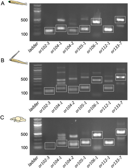
RT-PCR amplification of 7 olfactory receptor gene transcripts in non-olfactory tissues in larval and adult zebrafish. (A) or102-3, or104-1, or104-2, or105-1, or106-1, or112-1 and or133-7 genes are expressed in the whole 6-day-old zebrafish larvae. (B) All 7 OR gene transcripts are amplified in larvae without nose. 6-day-old Tg(OMP: YFP) larvae were used to ascertain the complete removal of the olfactory epithelium. (C) All tested OR gene transcripts are detected in the adult zebrafish brain. 100 bp ladder is used. Cartoons on left depict conditions for tissues used for RT-PCR. Green dot on rostral part of larva (A) represents OMP:YFP expressing olfactory epithelium, which was removed for (B). White boxes outline bands that correspond to the expected size. Original non-cropped images are in Fig. S3. |
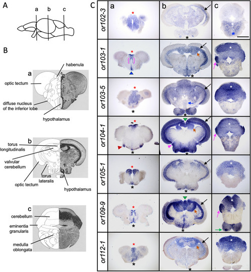
Olfactory receptor genes are expressed in the adult zebrafish brain. (A) Schematic drawing of a lateral view of the adult zebrafish brain indicating the anterior–posterior levels (a–c) of the transverse sections in (B) and (C). (B) Schematic image of transverse sections of adult zebrafish brain with different areas labeled in the forebrain (a), midbrain (b) and hindbrain (c) areas. Modified from Wulliman et al.24. (C) Representative transverse sections of adult zebrafish brains processed by in-situ hybridization for or102-3, or103-1, or103-5, or104-1, or105-1, or109-9 and or112-1 show different patterns of expression in distinct brain regions. Olfactory receptor gene transcripts are found in the habenula (red asterisk), preoptic area (blue arrowhead), hypothalamus (black asterisk), ventral thalamus (magenta bracket), diffuse nucleus of the inferior lobe (red arrowhead), optic tectum (black arrow), torus longitudinalis (green arrowhead), valvular cerebellum (orange arrow), eminentia granularis (magenta arrow), interpeduncular nucleus (blue arrow) cerebellum (white asterisk), medulla oblongata (blue asterisk), torus lateralis (magenta arrowhead). Scale bar = 500 μm. EXPRESSION / LABELING:
|

ZFIN is incorporating published figure images and captions as part of an ongoing project. Figures from some publications have not yet been curated, or are not available for display because of copyright restrictions. EXPRESSION / LABELING:
|
|
ZFIN is incorporating published figure images and captions as part of an ongoing project. Figures from some publications have not yet been curated, or are not available for display because of copyright restrictions. EXPRESSION / LABELING:
|