
Figure 2
|
Figure 2
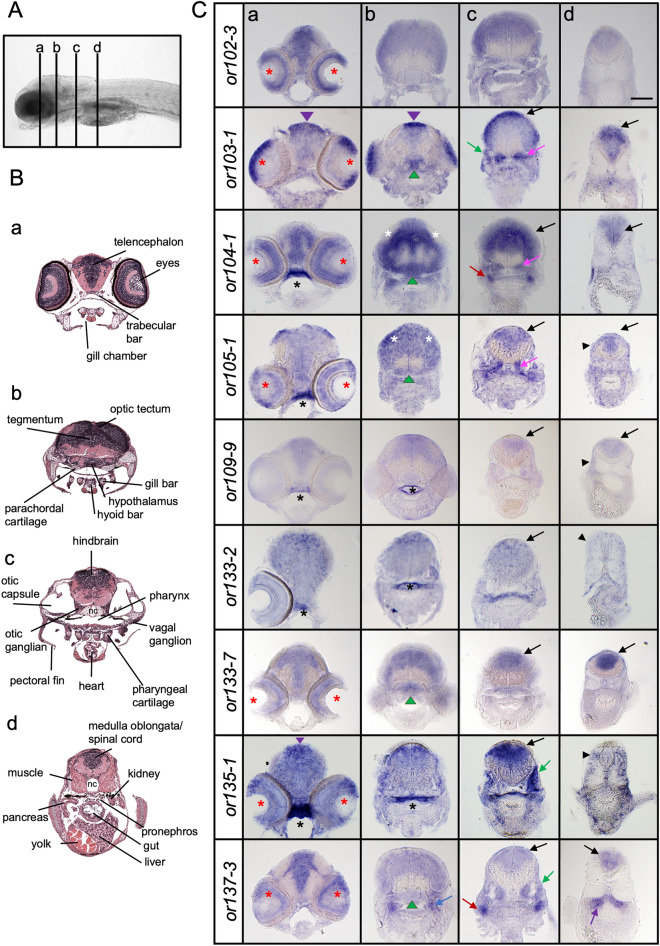
Olfactory receptor genes are expressed in distinct areas in the larval head. (A) Lateral view of a zebrafish larva showing the anterior–posterior levels (a–d) of the transverse sections in (B) and (C). (B) Images of transverse sections of 7-day-old larvae with different structures annotated. Sections represent the forebrain (a), midbrain (b), hindbrain (c) and posterior hindbrain (d). Modified from Bio-Atlas (Jake Gittlen Laboratories for Cancer Research). (C) Transverse sections of 6–7-day-old zebrafish larvae processed by in situ hybridization showing expression of olfactory receptor genes in different areas of the head. The stainings are localized in the dorsal midline (purple arrowhead), eyes (red asterisk), roof of pharynx (black asterisk), optic tectum (white asterisk), hypothalamus (green arrowhead), otic capsule (green arrow), otic ganglion (pink arrow), anterior lateral line ganglion (blue arrow), pronephros (purple arrow), vagal ganglion (red arrow), hindbrain (black arrow) and muscle (black arrowhead). Scale bar = 100 μm.