FIGURE SUMMARY
Title

Evaluation of a Rapid and Simple Method for Assessing Retinal Vessel Structures in Adult Zebrafish

Authors
Lee, Y.R., Son, M., Kim, Y.S., Kim, J.S., Kim, C.H., Jung, S.H.
Source
Full text @ Int. J. Mol. Sci.

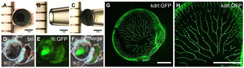
A rapid and simple method for the separation of the intact retinal vasculature from adult zebrafish eyes. (AF) The simple procedure of detaching the whole retinal vasculature from a dissected eye using a 1 mL micropipette tip. After the removal of the lens, the intact retinal vasculature was examined by high-resolution microscopy. (G,H) Fluorescent image of whole retinal vasculature separated from adult transgenic zebrafish line Tg[kdrl:EGFP], showing fine blood vessel structures. Scale bars; 1 mm for (AF) and 200 μm for (G,H).

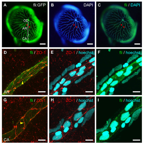
The cellular structure of whole-mount retinal vessels isolated from adult transgenic zebrafish, Tg[fli1:EGFP]. The vessel-specific expression of GFP (A) and nuclear staining with DAPI (B) in the retinal vasculature. (C) Merged image of (A,B). Retinal vessels from the optic disc (OD) branched out into the arterial region (AR) and then the capillary area (CA). Asterisks indicate membrane regions between blood vessels. (DI) Immunofluorescent labeling of tight junction protein (ZO-1, red), nuclear staining (Hoechst, blue), and GFP expression (green) in different retinal vessel regions; arterial region (DF) and capillary area (GI). Endothelial cells are elongated and their long axis is parallel to the line of the blood vessel. Arrows show cellular junctions between endothelial cells. Scale bars; 150 μm for (AC), 20 μm for (DF), and 10 μm for (GI).

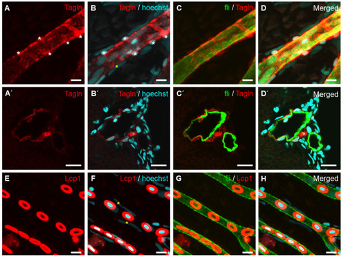
Identification of vascular smooth muscle cells in a retinal vessel of adult Tg[fli1:EGFP] zebrafish. (AD’) Vascular smooth muscle cells stained with anti-Transgelin (red) are distinguishable from GFP-expressing endothelial cells (C,C’; green). (AD) Whole-mount retinal vessel. (A’D’) Transverse section of the eye. Cellular nuclei were stained with Hoechst 33342 (blue). (EH) Hematopoietic cells were detected with anti-Lcp1 antibody staining (red) in the capillary region of the retinal vasculature. Asterisks indicate perivascular cells (A,B,F). Scale bars; 10 μm for (AD’) and 5 μm for (EH).

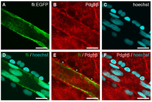
Identification of pericytes in retinal vessel structures. Confocal fluorescence image of vessel stained with anti-Pdgfrβ (B,E,F; red) and Hoechst 33342 (C,D,F; blue). Merged image of (A,C,D), (A,B,E), or (B,C,F), respectively. Vascular smooth muscle cells are Pdgfrβ-positive and surrounding vascular endothelial cells. Pdgfrβ-positive pericytes (asterisks) are also distinguishable from vascular endothelial cells (DF). Scale bar, 20 μm.

Changes in the retinal vasculature in high glucose-treated adult zebrafish. Vessel diameter was increased in high glucose-treated zebrafish (A’E’) compared to the control (AE). Confocal fluorescent image of vascular GFP expression (B,B’,C,C’), anti-ZO-1 (D,D’), or anti-Transgelin (E,E’) antibody staining. Arrows indicate retinal vessels. Arrowheads indicate representative cells stained with the specific antibody. Scale bars, 200 μm for (A,A’), 100 μm for (B,B’), and 50 μm for (CE) and (C’E’).

Acknowledgments
This image is the copyrighted work of the attributed author or publisher, and ZFIN has permission only to display this image to its users. Additional permissions should be obtained from the applicable author or publisher of the image. Full text @ Int. J. Mol. Sci.