IMAGE

Figure 3

ID
ZDB-IMAGE-221214-234
Source
Figures for Lee et al., 2022
Image
Figure Caption

Figure 3

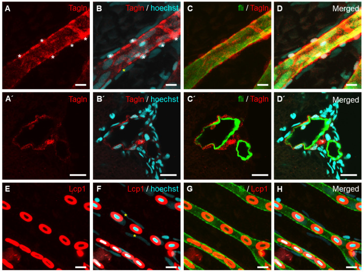
Identification of vascular smooth muscle cells in a retinal vessel of adult Tg[fli1:EGFP] zebrafish. (AD’) Vascular smooth muscle cells stained with anti-Transgelin (red) are distinguishable from GFP-expressing endothelial cells (C,C’; green). (AD) Whole-mount retinal vessel. (A’D’) Transverse section of the eye. Cellular nuclei were stained with Hoechst 33342 (blue). (EH) Hematopoietic cells were detected with anti-Lcp1 antibody staining (red) in the capillary region of the retinal vasculature. Asterisks indicate perivascular cells (A,B,F). Scale bars; 10 μm for (AD’) and 5 μm for (EH).

Acknowledgments
This image is the copyrighted work of the attributed author or publisher, and ZFIN has permission only to display this image to its users. Additional permissions should be obtained from the applicable author or publisher of the image. Full text @ Int. J. Mol. Sci.