
- Title
-
Essential Roles of the Histone Demethylase KDM4C in Renal Development and Acute Kidney Injury
- Authors
- Pan, H.C., Chen, Y.H., Fang, W.C., Wu, V.C., Sun, C.Y.
- Source
- Full text @ Int. J. Mol. Sci.
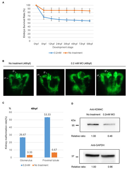
Embryo survival and kidney phenotypes of zebrafish embryos (96 untreated embryos and 96 embryos treated with kdm4c-MO injection). (A). Comparison of embryo survival rate between embryos treated with a single injection of 0.2 mM kdm4c-MO and untreated embryos (n = 96 in each group). (B). Pronephric phenotypes of zebrafish embryos after kdm4c-MO injection. Each kidney photo was taken from the dorsal view and at the developmental stage of 48 hpf of bTg(wt1b:EGFP) embryos. Compared with the untreated group, after kdm4c-MO injection, the renal tubules were tortuously dilated and the glomeruli were swollen. (C). Comparison of the defect rates of morphological phenotypes (tortuously dilated renal tubules and swollen glomeruli) of zebrafish embryos between untreated group and kdm4c-MO injection group. (D). Results of Western blotting for Kdm4c with zebrafish embryo kidneys with or without kdm4c-MO injection. Abbreviation: KDM4C, Lysine demethylase 4C; MO, morpholino oligonucleotides. EXPRESSION / LABELING:
PHENOTYPE:
|

KDM4C inhibition increased cell apoptosis during oxidative stress in vitro. HEK293 cells treated with the KDM4C inhibitor, JIB04 (1 μM) were used for the study. H2O2 (100 μM) treatment was used to induce oxidative stress. Cell apoptosis was analyzed by flow cytometry at the time points indicated in the figure. The percentages of dead cells at different time points after treatment are shown. Abbreviation: KDM4C, Lysine demethylase 4C. |
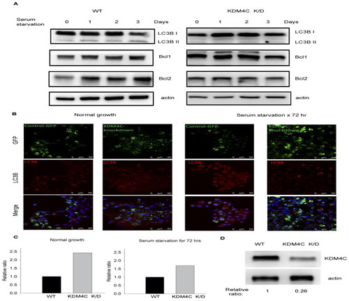
KDM4C depletion impaired autophagy during serum starvation in vitro. HEK293 cells transfected with KDM4C-sh plasmid were used for the study. Cells were harvested after serum starvation at the time points indicated in the figure. (A). Results of Western blotting for autophagy markers, LC3B, BCL1, and BCL2. (B). Results of immunofluorescence staining for LC3B in control and KDM4C knockdown HEK293 cells with or without serum starvation. (C). Relative ratio of fluorescence intensities of GFP-LC3B puncta with WT as the denominator. (D). Results of Western blotting for KDM4C in control and KDM4C knockdown cells. Abbreviation: K/D, knock down; KDM4C, Lysine demethylase 4C; WT, wild-type. |
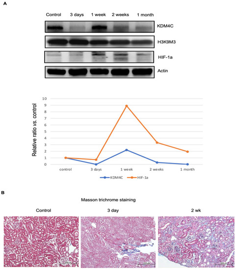
Temporal changes in KDM4C and progression of kidney fibrosis during kidney injury. Wild-type mice receiving kidney ischemia-reperfusion injury (IRI) were used for this study. Kidney tissues, after IRI for the durations indicated in the figure, were harvested (n = 8 for each group). (A). Western blotting results for KDM4C, H3K9M3, and HIF-1α. The average relative ratios of KDM4C and HIF-1α along the course of injury are shown. (B). Masson trichrome staining results of kidney tissues after IRI. Mild glomerulous and tubulointerstitial fibrosis, 3 days after receiving IRI-AKI. The severity of glomerulous and tubulointerstitial fibrosis was even more pronounced 2 weeks after IRI-AKI. Abbreviation: HIF, hypoxia-inducible factor; KDM4C, Lysine demethylase 4C. |

KDM4C depletion worsened acute kidney injury. Study animals receiving kidney ischemia-reperfusion injury were used for this study. Kidney tissues and serum after ischemia-reperfusion injury for the durations indicated in the figure were harvested (n = 8 for each group). (A). Serum BUN and creatinine levels of wild-type and Kdm4c−/− mice after kidney ischemia-reperfusion injury. (B). Masson trichrome staining results of kidney tissues of wild-type and Kdm4c−/− mice after ischemia-reperfusion injury. (C). Representative results of kidney inflammatory cytokines of the study animals, 1 week after ischemia-reperfusion injury. (D). Results of Western blotting for KDM4C in the kidneys of wild-type and Kdm4c−/− mice at the age of 10 weeks. Abbreviation: BUN, blood urea nitrogen; CCL12, C-C motif ligand 12; CXCL1, C-X-C motif ligand 1; FGF, fibroblast growth factor; IGFBP-2, insulin-like growth factor binding protein-2; IL-1R, interleukin-1 receptor; KDM4C, lysine demethylase 4C. KIM-1, kidney injury molecule-1; NGAL, neutrophil gelatinase-associated lipocalin; WT, wide-type. * p < 0.05. |
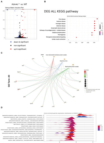
Differential kidney expressions of genes between the wild-type and Kdm4c−/− mice. Kidney tissues of wild-type and Kdm4c−/− mice at the age of 10 weeks were used for this study (n = 8 in each group). (A). Volcano plot showing differential kidney gene expressions between the wild-type and Kdm4c−/− mice. The results showed that 42 genes had higher expression levels, while 46 genes had lower expression levels in the Kdm4c−/− mice than in the wild-type mice (p-value < 0.01, q-value < 0.01). (B). The KEGG pathway analysis results showed that Kdm4c deletion was also associated with a variety of neurodegenerative diseases and metabolic diseases. (C). GO enrichment analysis showed that the Kdm4c−/− mice had significantly different expressions in several pathways, including inner mitochondrial membrane protein complex and mitochondrial protein complex. (D). GSEA revealed that Kdm4c deletion was also associated with several pathways involved in mitochondrial function, including mitochondrial electron transport chain, oxidative phosphorylation, and mitochondrial membrane phospholipid-based signal pathways. Abbreviation: KDM4C, lysine demethylase 4C; KEGG, Kyoto Encyclopedia of Genes and Genomes; GSEA, gene set enrichment analysis; WT, wild-type. |